Introduction
Idiopathic intracranial hypertension (IIH) is a disorder that leads to isolated raised intracranial pressure characterized by classical symptoms and signs such as headache, papilledema, sixth nerve palsy causing diplopia and pulsatile tinnitus.1 Papilledema is the most frequently encountered clinical finding in IIH.2 Our case presents a 20-year-old primigravida with unusual signs of bilateral proptosis, unilateral ophthalmoplegia, and unilateral facial nerve palsy. In previous literature, there are only two reported cases of IIH that were associated with proptosis, both presenting unilaterally, and one reported case of IIH presenting with complete unilateral facial palsy. 3, 4, 5
Case History
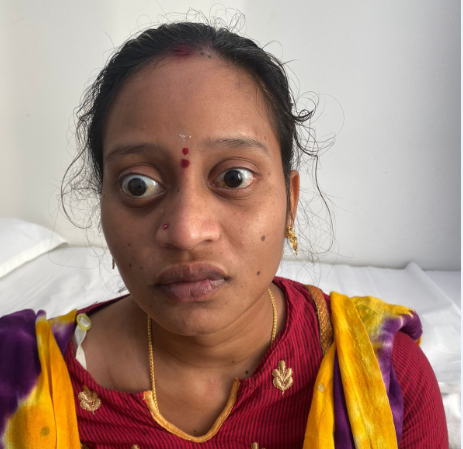
Twenty-year-old female, a primigravida with a gestational age of 23 weeks, presented with complaints of right temporal headache for the past ten days and cervical pain for the past three days which was radiating to bilateral supraclavicular region and the right upper limb. History of decreased vision in both eyes for five days, diplopia for three days associated with ringing sensation in bilateral ears, and protrusion of both eyes were also present. On examination, she was found to have bilateral proptosis (right > left) (Figure 1), impaired upgaze, adduction and abduction of the right eye, and right seventh CN lower motor neuron palsy (Figure 2). The proptosis was axial and non-pulsatile. Her visual acuity (VA) was 20/20 OU and color vision was intact. Both pupils were equal and reactive to light. Baseline investigations (complete blood picture, renal function tests, liver function tests, hormone levels) were found to be normal. Based on the clinical profile, possible diagnoses considered were: 1). thyroid ophthalmoplegia, 2). idiopathic intracranial hypertension, 3). bilateral cavernous sinus thrombosis. Imaging with an MRI brain + venogram was suggestive of vertical kinking of the right optic nerve and tortuosity of bilateral optic nerves and prominent bilateral perioptic cerebrospinal fluid (CSF) spaces along with mild indentation or flattening of the posterior sclera (right > left) (Figure 3). It also revealed stenosis in bilateral transverse sinuses (Figure 4), and no evidence of thyroid ophthalmopathy or cavernous sinus involvement. Lumbar puncture was done to detect the opening pressure of CSF which was measured to be 57cm of water. CSF analysis and cell counts were not indicative of any pathology. Fundoscopy revealed bilateral swollen, hyperemic, enlarged optic discs with blurred margins suggestive of bilateral papilledema (Figure 5). Visual fields revealed enlarged blind spots in both eyes. She was managed with 250 milligrams of acetazolamide twice daily which was continued on discharge. After the course of medication, the patient experienced alleviation of symptoms along with a significant reduction in proptosis bilaterally and restored extraocular movements of the right eye.
Discussion
The etiology of IIH has been debated in literature so far but a definitive cause still seems to be elusive. 6 There are several proposed hypotheses, most of which revolve around CSF homeostasis, including surplus CSF production, decreased CSF absorption, diffuse brain edema, and increased cerebral venous pressure. 6, 7 It has been proposed that diagnostic criteria for IIH should comprise a lumbar opening pressure greater than 250 mm of water measured with the patient in the lateral decubitus position. 8 Prevalence of IIH is highest amongst obese women of child-bearing age. 9
Some unusual or atypical presentations of IIH include ocular motor disturbances from third nerve palsy, fourth nerve palsy, internuclear ophthalmoplegia, and diffuse ophthalmoplegia; unilateral papilledema or IIH without papilledema; olfactory dysfunction; trigeminal nerve dysfunction; facial nerve dysfunction; hearing loss and vestibular dysfunction; lower cranial nerve dysfunction including deviated uvula, torticollis, and tongue weakness; spontaneous skull base cerebrospinal fluid leak; and seizures.1 In previous literature, there are only two reported cases of IIH that were associated with proptosis, both presenting unilaterally.3, 4 Out of the two, the first one is that of a 24-year-old woman who presented with a history of visual abnormality and was later discovered to have bilateral optic disc swellings, dilatation of the optic nerve sheaths, and increased CSF pressures. A diagnosis of dural ectasia was considered. Usually, papilledema and optic nerve sheath enlargement can be seen with IIH. 4 However, notable evidence suggesting the role of raised intracranial pressure in the etiology of dural ectasia of the optic nerve is lacking. 7, 10 The latter reports a 22-year-old female presenting with headache with vision loss, papilledema, complete ophthalmoplegia with proptosis in one eye, and sixth CN palsy in the other eye. 3 In our case, we notice predominant cervical pain, bilateral proptosis, external ophthalmoplegia, and right seventh CN palsy.
The most common CN palsy seen in IIH is that of CN VI, and less frequently, other CNs are involved like CN III, IV, VII, IX, and XII. In 2019, Ahmad Samara et al reported a case of IIH in which an obese and hypertensive 40-year-old Hispanic woman presented with isolated complete unilateral facial nerve palsy. Her neurologic examination was otherwise normal, but a fundus examination revealed bilateral papilledema. The precise pathogenesis behind facial nerve involvement is not entirely understood. However, it is widely described as a false localizing sign resulting from increased ICP exerting traction forces on the extra-axial facial nerve. 5
Transverse sinus stenosis (TSS) is a common finding in patients suffering from IIH, detected on neuroimaging. It probably contributes to rise in ICP via venous hypertension. 11 It is almost always bilateral which impedes cerebral venous outflow to a significant extent. 11, 12
Various methods of management have been proposed for IIH. A conservative approach would be to focus on strategic weight loss. Medication usage includes the administration of diuretics. For those cases that are refractory to conservative management, surgical intervention might prove to be imperative. Surgical options include CSF diversion or optic nerve sheath fenestration, venous sinus stenting, and bariatric surgery. 13
Conclusion
After reviewing previous literature, we were able to find very few reported cases of IIH that were associated with bilateral proptosis, unilateral ophthalmoplegia, and unilateral facial nerve palsy together and this is the first case of IIH to be reported with this unconventional presentation. When a patient presents with these symptoms and signs, the diagnosis of IIH cannot be excluded. The cause of bilateral proptosis can be postulated to be attributed to the rapid increase in ICP, the magnitude of elevated ICP, pregnancy, and its hormonal influence. As the patient’s symptoms were relieved after administration of acetazolamide, the hypothesis stands true. Further studies are warranted regarding cases of IIH with unusual presentations as witnessed in our case report.


