Introduction
Out of the excessive efforts, many years of hard work with dedication and long experimental research by two pioneering neuroscientists’ namely, Louis Benabid1 and MeLon DeLong2 from their exploratory labs, the development of deep brain stimulation (DBS) was born, and it is now offered at many DBS clinical centers worldwide and offering better solutions as a therapeutic benefit to the Parkinson`s diseased subjects, i.e., patients.3 Hence, a huge applause to the duo scientists and the human beings have had entered the era of humanoid-neuronal/ or neural networks modulation.
DeLong4 formulated a new model for the brain's circuitry and exposed a fresh target for this illness. Benabid5 devised an effective and reversible intervention that remedies neuronal misfirings. Their work has culminated in an effective treatment for more than 2,50,000 individuals worldwide with severe illness who suffer from complications of levodopa therapy.6
The signals of STN neurons acquired with intra operative MER might contain vital data and info regarding - in relation to functional activities of brain region-structures which could be correlated in conjunction with disease-symptoms.5, 6, 7 Data indicates that stimulator (the DBS) ensures not only impacts neural-tissue at the point-of-electrodes (PoE) however disrupts pathological-signals that resonate across several brain-regions. Gathering LFPs by SVM-based MER signals recording from STN neurons with DBS lead can give valuable feedback for titrating therapeutic DBS technique and for examining the neural network modulation mechanism of the DBS therapy, else, in the investigation of adaptive closed-loop BDS-devices, the method yielding continuous high-frequency induced electrical stimulus usually employed in clinical-diagnosis practice. Titration is a procedure or process to quantify a substance in a solution, a common technique employed in analytic-systematic critical-diagnostic methodical-chemistry to define or ascertain the strength/concentration of an unidentified or not known resolution thru progressively accumulating a solution (resolution) by an identified absorption (concentration). The reactance of recognized intensity (i.e., concentration) is improved tad by tad and bit by bit till nullification (neutralization or offsetting) is attained.
Even if structural organization, i.e., anatomical structure of an organ of the brain gives a few clues or pointers as to what might be the function of basal ganglia circuits in Parkinson`s, albeit the inference or implicational extrapolations or reason of function from anatomical structure is exploratory. Therefore, experimental investigations for the objective evidence are necessary. So, one investigative approach to studying-the-function of an area-of the central nervous system (CNS) substantia-nigra (SN) is to acquire the STN neurons with extracellular MER in locally anesthetized PD patients.8, 9, 10, 11 Other approaches involve inferences of neuronal signaling from imaging studies of blood flow and metabolism, or of changes in gene-expression. By sampling the signal of a part of the brain during behavior, one can gain some insight into what role that part might play in behavior. Neurons within different basal ganglia nuclei have characteristic baseline discharge patterns that change with movement.12, 13
Because of the aging, there is a every possibility to increase the brain diseases and disorders and there by symptoms chances are more and more. Hence, examining the intra operative (intra op) electro physiological activities with acquiring with microelectrodes and stimulations via DBS device plus how the stimuli impact the behavior and the behavioral actions and activities of the brain during the DBS process is imperative.
Though, the stimulus intensity, i.e., amplitudes of the stimulus pulse-widths are very huge contrasted to the neuronal activities (neural actions), making the concurrent sensing during the stimulation difficult. It is full of challenges to inhibit the strong stimulation pulses as well as reserving the full information of the neural activities. In this connection, the active personal computer plus system offering an implantable device for acquiring the data parallelly by sensing the STN or pallidal neurons with DBS stimulators as a research tool to investigate the neural-circuits during the therapeutic DBS procedure and the process is very innovative. This has given some understandings of PD brain activities functionally. 14, 15
Earlier research demonstrated the design of an implantable electroencephalography(EEG) device-based on with high rich skill developing DBS devices which not only offers the therapies but then again likewise yields better parameter measurements as a basic tool for the biomedical neuroengineering signal processing applications to Parkinson`s disease and other movement disorders in neuroscience.16 But, few residual stimulus-artifacts are occurring in the signals acquired while the stimulus DBS is ON (i.e., “DBS-ON or ON-DBS” and the medication is off, i.e., “MED-OFF”) which is inexorable or inevitable as a result of owing to the limitation of the implantable-device, which includes accumulation (storage capacity) plus power consumption. Thus, it is necessary to restrain the residual stimulus device artifacts occurring due to the stimulations by DBS and there by lacuna in the data-processing.
The artifacts due to the stimulations by DBS normally induce phantom-spectra, i.e., spectral noise by the stimulus-frequency and thereby its harmonics or oscillations or ripples and this is occurring mainly due to the improper elimination of aliasing by sampling (through the filters at the hardware design and development and hardware programming levels) and improper windowing to minimize the spectra (at the processing level, i.e., software programming levels). Also, while devising the devices perfect filters are not employed or perfect filters are not designed (prior to development of the filter-devices). Many techniques were employed by the many researchers previously to repress artifacts during DBS in electro-neuro-physiological signals acquired through continuous DBS stimulations with microelectrode recording machine.16 Some filters like notch or low pass are basic solutions. However, if the induced stimulus-peaks overlap numerous frequency-bins, then such filters are inadequate and inefficient and not suitable.17 Some researchers focused on addressing the suppression of DBS artifacts on the templates model abstraction-techniques. 18 But the template abstraction-techniques endure as of the notions that the phasal (shapes of the waveform or signals) artifacts are continuous and in steady-state amongst stimulus-pulses. Therefore, these techniques are frequently applied in extracellular signal acquisitions by sampling-rates at higher to avoid higher frequencies components noise and distortions. Researchers’18 built an artifact-template (with stimulations) by window-averaging technique throughout the peri-stimulus-segments. But the sampling-frequency of analog to digital converter (A/D C) conversion modules in embedded-systems (implanted-devices) generally won’t exceed beyond the 1k Hz due to low-power constraints of implantable-medical-devices. Such as, the Activa PC-based DBS system employed nearly ~ 423 (≤422Hertz) in which every stimulus-artifact include quite of few numbers of sampling-points with kinds of morphological data followed by the model-template subtraction-technique with which it may be difficult to process or execute and finally difficult for implementations.
In this study, based on stimulus-artifact re-concentration plus elimination, we proposed a novel-technique to eliminate the artifacts in the MER signals of STN neurons and extract LFPs without any unwanted signal-noise components while keeping the STN neural signals in LFPs acquired during parallel stimulations through deep brain stimulator. The method is evaluated in-vitro experimental tests using the mock electrical potentials of local fields by induced stimulus and devoid of stimuli to differentiate suppressed artifacts. The method is examined in both in-vitro and in-vivo.
The method is then used to process the LFPs that were recorded in-vivo from the STN of PD patients during DBS surgery, with the signal characteristics dependent on the stimulation parameters.
Aims and Objectives
The objective is to study the correlation amongst the subthalamic-nucleus (STN) deep brain stimulation (DBS) parameters and the variant of STN-LFPs. To minimize the total-elapsed time for the duration of the surgical DBS operational procedure, an (automatic) pre-programed stimulus-charging program is employed to automatically adjust the induced stimulus-parameters. To propose a method to suppress the residual-noise of deep brain stimulation (DBS) stimulus-artifacts throughout the data processing.
Materials and Methods
The technical configurations, specifications and performance of the instrument influence the accuracy of measurement such that meaningful recording of electrical activity of the brain can only performed with properly designed equipment.
The range of voltage and frequency encountered through microelectrode recording (MER) of STN neurons is far too less than that observed in muscles electromyography signals recorded through Electromyograph (EMG), electromyography signals recorded through electroencephalograph (EEG), and electrocardiography signals or cardiac signals acquired through Electrocardiograph (ECG), machines. The objective in recording is to obtain a faithful reproduction of the physiological event, i.e., signals picked up by the electrodes should be amplified and presented to the examiner/ user–observer without noise – distortion and free from any interference which may obscure the signal.
A. Design, development, and application of instrument
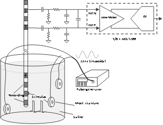
The condensed-void amongst the five microelectrodes (4 reference, and 1 ground) plus the giant amplitudes of the electrical-pulses (generated by the implanted pulse-generators called IPGs), order of the magnitudes typically 5 to 6 higher than that of the electric-field-potentials (EFPs) round the microelectrode, made it problematic to acquire LFPS parallelly throughout the stimulations by DBS. Earlier researchers19, 14, 20 were applied the proportional or symmetrical proportionate and equally balanced geometry-of-electrode [#VRR IETE] to gather differential-field potentials, i.e., local-field electric-potentials amidst adjacent neighbour-microelectrodes on the identical-lead in conjunction with unipolar-stimulus.[9,18] Through this microelectrode’s technical-specification based on benchmark formalism, the distance from the implanted pulse generators (IPGs) to the DBS lead (macroelectrode) is considerably larger than the space amongst the DBS stimulus-electrodes. Thus, the due or twosome acquiring-electrodes, the two of a kind will sense involving the identical stimulus-pulses. And the then stimulus-interference be seen as a common-mode-rejection (CMR) disturbance to the input differential-amplifier. The same symmetrical geometry-of the electrode concept is employed in this study and is depicted in the pictorial representation as a schematic diagram Figure 1.
A monopolar-stimulus-mode (MSM) applied in conjunction with a titanium-stimulator-case (TSC) as an anode-electrode, i.e., passive or positive charge +Ve electrode (the terminal or electrode from which electrons leave a system) with a passive terminal, i.e., passive or positive (+Ve) electrode and one of the two middle contacts as cathode-electrode, i.e., active negative (-Ve) electrode (while charging, the passive, i.e., +Ve electrode is an anode, and the negative electrode, i.e., active, i.e., -Ve is a cathode). The -Ve charge electrode functioned as the ground-electrode for sensing-module of local field potentials. Bipolar stimulus charging electrodes were employed by some studies previously21, 20, 22, 23 during the evoked local field potentials generated with the DBS by applying the stimulus-pulses amid 4(positive, i.e., +)electrodes and 2(negative, i.e., -Ve) charging electrodes during the generation of differential local field potentials among the charging electrodes 1 plus 3. Even though this nonsymmetrized (asymmetrical) electrode structure and arrangement hypothetically intervene higher-noise into the generated data, it’s possible to observed real-signal (by the observer) and reinstate and reproduces at the processing level effectively as discussed below (provided the A/D converter supports required dynamic range, adequate sampling frequency and the sampling conversion time of the A/D dynamically):
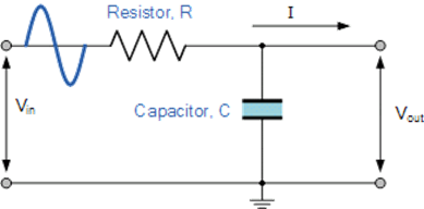
A simple but elegant low-passive filter (Figure 1, Figure 2) network with a pass band of 0.2Hz to 100.0Hz suppressed the larger-amplitudes (from 1volt to 10volts signals) to merely some 100μ-Volts in the common-mode-rejection (CMR) input range of the A/D converter.24
The Texas Instruments ADS1257 24-Bit Analog-to-Digital Converter (ADC) is a low-noise, 30kSPS, 24-bit, delta-sigma (ΔΣ) analog-to-digital converter (ADC) is a programmable and front end analog 24 bit and back end 24 bit digital for biomedical applications especially for measuring the bio medical signals or potentials and bio medical and biological signals applications which consists of 8 channels of ∆-∑ A/D converters. A/D converter details are given at the Appendix.
The basic internal-cells of ∆-∑ A/D converters in the A/D converter is the ∆-∑ modulator through higher frequency-modulation on 1.024 Mega Hz plus the digital decimation or annihilation filter, which is, a third-order low-pass sync-filter by a changing annihilation rate. Also, the on-chip decimation-filters offer anti-aliasing-filtering (AAF), i.e., for eliminating the ripples or oscillations as well as filtering out high frequency-noise. The AA filter is applied prior to the sampling of the signal to impede the bandwidth of a signal to fulfill the “Nyquist–criteria and Shannon`s communication” sampling-theorem over the band-of-interest.
The module for the sensing of the local field potentials is designed to be integrated into the rechargeable implanted pulse generators (IPGs), i.e., neurostimulators through the bilateral subthalamic-nucleus deep brain stimulation pulses output. The radio frequency signals, which travel by using a variety of movement behaviors referred to as the propagation-behaviors. The acquired data shall be done by employing the wireless radiofrequency (RF) communiqué message in real-time simultaneously (the amount of processing that can be accomplished in the given interval of time). The recharging via wireless can be the trade-off towards the power-consumption additionally for the field potentials gathering as well as broadcast communication-transmission/functions.
Maximum acquisition time through this system is till 4 to 5 hours plus the 2 channel signals with a sampling frequency range of 1kHz on a specific singular battery-charge.
B. Experimental Set Up
In this study, we have setup for a real-time multi-channel capturing of local field potentials with special reference to Parkinson`s from the subject having input impedance greater than 300 Mega�𝝮. An input-impedance of at least 10Mega�𝝮 is required for amplification system, CMRR of 65 dB and variable gain 1000-10000. Ag electrodes used. Good differential amplifiers with CMRR should have rejection ratio close to 100,000. In other words, the signals are amplified 100,000 times more than unwanted potentials appearing as a common mode voltage. The input impedances of the most amplifiers range from 100 k𝝮 to hundreds of Mega�𝝮s.
In-vitro setup, the method is tested to produce the constraints of stimulus-acquisition with the DBS using the implanted pulse generators - microelectrodes within the STN region of the PD brain, as shown in Figure 2. The DBS having 4 platinum–iridium cylindrical-contacts with a 1.3mm diameter followed with a length of 1.5mm, and spaced at 0.5mm apart, then limit a complete distance of 7.5mm), come to 1 to 4 from the best caudal to the highest-rostral-contact(HRC). A Pins L301 DBS lead is immersed into a glass container filled with saline solution (9gms of sodium-clorideNaCl/liter-water) at room temp and is applied to provide mono-polar-stimulus (MPS) plus to acquire the signals among the adjacent non-stimulating-contacts. Two additional Ag/AgCl disc-electrodes which were usually used for recording scalp EEG were used to inject sinusoidal simulated LFP generated by a function (purse and/or signal) generator into the saline-solution. Given that the frequency spectrum of
A custom program written in visual C++(B-Tree binary tree implementation efficient for arithmetic operations and a graphical tool for the visualizations) operated the elements in the signal-data acquisition-module to set up the digital target-output timing followed by sampling frequency of the signal (default: 1kHz sampling-rate).
Figure 2
Schematic-diagram of in-vitro experimental-set-up employed to gather local field potentials in the course of deep brain stimulation.

A lead of deep brain stimulation is immersed in a saline-bath for acquiring and to provide stimuli. Two Ag-AgCl discoid-electrodes were set on either-side of DB-stimulator lead to produce the simulated-LFP (23Hz sinusoidal-signa pulse-generator).
C. Signal-chain discussion
With the symmetric electrode configuration illustrated in Figure 2, the two recording electrodes sense approximately the same stimulation pulses. After passing through the filter network, the input signals were suppressed. The digital decimation filter received the modulator output and decimated the data stream, which consisted of a third-order Sync filter with the Z-domain transfer function
|H(z)| =
Where, ‘N’ is the ratio of the decimation.
D. Removal of noise-artifacts
Stimulus artifacts-characterization
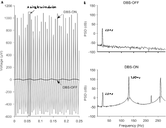
In most-of the cases, the stimulus-artifacts can be connected as a linear-stationary-signal (LSS). According to the Discrete-Fourier-Transform (DFT), the frequency-spectrum (of the power spectral density) issue comprises of harmonic-frequency-components (HFC) that are integer multipliers of the basic fundamental-frequency. As well as the harmonic-oscillations, potential analog to digital converter aliasing (ripples due to improper sampling by the A/D filters at the hardware levels) brings in aliased-artifact-frequencies (AAF)s which can be defined from the sampling-frequency. Therefore, the harmonic-oscillations (hOs) and aliased-artifacts (AAs) can be clearly and elegantly found for the given stimulus plus in sampling-frequencies. A signal acquired in the amplitude 2Volts, pulse-width 90μs, and frequency 130Hz stimuli in the saline-solution (SS) is depicted in Figure 2.A, also the signal acquired when the stimuli is OFF (i.e., DBS-OFF) is also shown, which worked as a reference-signal, i.e., as a good and observed signal. Figure 2.B shows the power-spectrum of the signal-acquired in induced-stimuli and that of the signal when stimuli is OFF, i.e., DBS-OFF. Because a 23Hz sinusoidal-signal is infused into the saline-solution to induce the local field potentials, this frequency is a peak at 23Hertz in the power-spectrum, i.e., power spectral density spectrum.
A. Segments-of0.25seconds of initial-time-signals acquired as of saline-bath when the stimulator is OFF, i.e., DBS-OFF (depicted as black-line), when DBS (Current-voltage:2Volts, frequency:130Hertz, pulse-width:90μs) is ON (grey line) and dashed-line-box signifies a single-stimulus-artifact(SSA).
B. The PSD of signal when DBS is OFF, i.e., DBS-OFF and the PSD-spectrum of the signal when DBS is ON, i.e., DBS-ON.
Noise removal procedure
As discussed in the above, the delta-sigma analog to digital converter modulated the differential-signals which are augmented at a higher-rate (1.024MegaHz) and then down-sampled to 1kHz following the anti-aliasing-filtering(AAF). Prior to down-sampling, the stimulus-artifacts of the filtered-signal were nearly similar, hence the basic-strategy is to rebuild a SSA of this stage by employing the de-trended raw-signal acquired to elicitate the SA-template by the separation plus superimposing of the every-artifact-noise.
Figure 4
A.Noise-removal (i.e., artifacts) through template-subtraction as ofinitial-original signal acquired from saline-bath in 180Hertz sinduced-stimulus.

A data sequence covering “M-number of data-points” from the de-trended ‘raw-data’ is employed for reconstructing the stimulus-artifact-template (SAT). This data sequence should circa ~ include an integral-number (indicated with ‘N’) of stimulus-pulse width noise-artifacts, which is defined as a result of evaluating the phases (shapes) of the first-stimulus pulse-artifact plus the one which is after this data-sequence. A linear-interpolation-method(LIM) is then applied to enhance the number-of data-points to the least-common-multiple (LCM)of ‘N’ and ‘M’ or ‘N’ times of ‘M’) This data sequence is then segmented into N segments with these segments-overlapping. lastly the down-sampled-template is subtracted from the raw-data to obtain required and imperative signal.
Artifact noise-removal procedure
Figure 4B. Demonstrates the SAR procedure. A data sequence from the de trended raw-data is applied for constructing the artifact-template. The “EMD-technique” is applied for de trending as discussed in the reference 17. This chosen data-sequence should have ~ an integral number-of-stimulus pulse-width-artifacts. The desired-artifact-shape (the phase) is then equated to phase-shapes of the continuing-artifacts sequentially to find alike one-by indexing one through min-average deviation-of artifact-point-value, i.e., to find the noise-artifacts convincing the following equation,
Where, ‘min’ remains for finding the minimum-value, and 𝑑𝑛 and 𝑑𝑛′ are the data-points of the two contrasted-artifacts, plus ‘n’ is the index-of the data-point of every-artifact. Therefore, we get a data-sequence that consists ~ an integral number-of stimulus-pulse-artifacts noise.
Mathematical evaluation
Following the removal-of-artifacts, the PSD of the recuperated-signal in the course of “DBS-ON” ought to be like to the PSD-spectrum of the signal when the stimulus is OFF, i.e., “DBS-OFF”. Which is estimated through the ratio ‘R’, which is stated in decibels “dB” which is the density of the spectra and computed using the following equation (3) as:
R =
Here, POFF(f) plus PON(f) indicates he PSD-spectrum of the signal while the stimulus is OFF, i.e., DBS-OFF and the retrieved-signal throughout stimulations i.e., DBS-ON at every-frequency ‘f’, and the mean ‘f’ relates to the arithmetic-mean (AM) over all frequencies. Several valuable local-field-potentials-biomarkers for D B S applications typically have frequencies below <= 100Hertz, for example the frequency-range for Parkinson brains which is the
And for in vivo-tests, ‘statistics’ on
Raw
Clinico-statistical analysis
The data is differentiated by applying the non-parametric Wilcoxon’s signed-rank- tests. The findings are registered as mean± standard-error of mean (‘S.E.M’). Pearson correlation-coefficient (P) is computed to estimate the connection amongst the stimulus-parameters followed by β-band control-level.
Results and Discussion
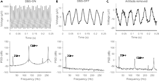
The segments of the signals acquired as of saline-H2O while stimulating, i.e., “DBS-ON” is contrasted through the segments while stimulating, i.e., “DBS” is “OFF” (DBS-OFF) in Figure 4A followed by in Figure 4B. The initial “DBS-ON” signal practically totally obfuscates the effective sinusoidal-signal since the huge stimulus-artifact-noise. But, the removed noise-artifacts yielded as sinusoidals (convenient for analytical purposes only but not efficient as a basis vectors like EMG, ECG, EEG, and MER signals) depicted in the Figure 4C., even though a few minor residual stimulus-artifacts persisted in the graphical-visual-inspection of the time series time-domain signals.
Figure 5
Segments-of0.25seconds of the initial-time-domain signals acquired as of saline-bath plus power-spectral-density, i.e., spectra of PSD of them.

A. Signal while DBS-ON (i.e.,”DBS-ON”) with setup-parameters: Voltage - 2V, Frequency - 130Hz, and pulse-width - 90μs.
Signal while the “DBS is OFF”, i e , DBS-OFF.
Signal following the removal of artifacts-noise.
Though earlier DBS devices could not record noise-free artifact-free signals, the Filter D B S19 can record the noise-free/artifact-free potentials while DBS is ON and it also minimizes the time-delay for computing with the bio signal i.e., biomarkers rejoinder to the stimuluses. The technique is applied to validate analogous-effects for example by dopamine chemical messenger that D B S microelectrode stimuli suppressed disproportionate and extreme
Conclusion
We proposed a novel method for removing the stimulus-artifact’s noise in the LFP signals, offering offline signal-processing of the PD data for the D B S devices through the function of sensing-neural electrical-activity for instance local field potentials while delivering DBS-therapy. The proposed method offers technical support for exploring the instantaneous effect of electrically induced-stimuli on the brain-activities of neurological disorders.


