Introduction
Micro electrode recording (MER) signals are enormously curve data. Most bio signals are referred to as biomarkers i.e., “bio signals”. These signals are assisting the clinicians to study the diagnosis and then for the findings the best remedy and best treatment. Further, acquired signals are guiding to develop the frontier technologies in the medical devices sectors. The machines like magnetic resonance imaging (MRI), Computed axial topography`s (CATs), transcranial magnetic stimulators, positron emitted tomography`s (PETs), deep brain stimulators (DBS), Micro Electrode recording (MER) machines were built based on the sample signals obtained from both the diseased subjects and from normal health controls helping highly to the development of cutting-edge frontier-technologies’ especially in health care sectors medical devices and systems for clear understanding of the disease and predicting symptoms of the disease in early stage. Such signals are modeled and designed by scientists and engineers (trans-disciplined i.e., i.e., scientists with diverse background from multi-disciplinary subjects).1, 2, 3, 4
The Parkinson disease is correlated in conjunction with numerous inconsistencies, i.e., abnormalities in the function of the brain as well as anatomical-structural arrangement. The pathological characteristic feature of the disease is a endless malfunction of nigro-striatal dopamine cells and an accumulation of intra cellular inclusions called “Lewy body`s” which are contained α - synuclein that gathers masses. Infact through dopamine- energic cell loss, a number of indeed numerous additional neuro transmitter systems as well degenerate and afterwards on “Lewy-body-pathology” also extends to the motor cortex. This creates Parkinson`s classic systems at structural-disorder, and this might be totally implicit and simply after examining the molecular empirically, anatomical-structure and practical inconsistencies and irregularities at the networked brain levels. Thus, the quest for ideal antidote, best cure, and best remedy is on for the past two centuries since the time it was first detected and described by James Parkinson in his research article published in a England journal.5 Nevertheless, scientists confirmed that the malady develops from inadequate amounts of the neuro transmitter dopamine (a chemical messenger) in an essential part of the brain that controls movement, the parallelly connected basal-ganglia(BG) circuits.6
One of the most effective treatments for PD is deep brain stimulation (DBS) of the subthalamic nucleus (STN). The procedure involves the surgical implantation of stimulating electrodes into the STN and provides a unique opportunity to record in vivo the related neuronal activity, through microelectrode recordings (MERs) of high spatio—temporal resolution. However, the optimal placement of the stimulation electrode continues to be a challenge, possibly due to the neuroanatomic variability within the STN sensorimotor area.7 MERs have been used before to enhance our understanding of how STN neurons function and classify possible mechanisms for DBS in PD. MER-based algorithms have also been developed to categorize and detect the sensorimotor area within the STN by using both the high and low-frequency content of the recorded signals.8
The high frequencies of the MER signal include both the electrical action potentials from neurons located closest to the electrode tip (typically at a distance of less than 100–300 μm) as well as smaller sub-noise level spikes from nearby neurons known as “background-unit-activity” designated as BUA. The combinations of these two signals are referred to as “multiunit-activity” referred to as MUA. The lower frequencies of the MER signal correspond to the local field potential called LFP potentials, which reflect the cumulative activity of a population of neurons within a better diameter from the electrode tip (around 0.5mm–3mm). So far, most of the MER based analyses of the STN have focused by a series of scientists7, 9, 10, 11, 12, 13, 14, 15, 16, 17, 18, 19, 20, 21, 8 on the gross automatic detection of the STN borders.
Amir novin performed the research on micro electrode signal recordings (MER) in objecting sub thalamic nucleus (S.T.N.) in 40 Parkinson`s disease candidates (i.e.,Parkinson diseased subjects). The expected site (with the pre operative magnetic resonance imaging MRI, and interventional STN-DBS, and GPi,e,) was employed in 42% of the cases. Nonetheless, in the left over 58%of the cases it was controlled all through the MER (i.e.,M.E.R by means of S.T.N-D.B.S). By applying the support-vector-machine (SVM)-based M.E.R method, an average pass-through the subthalamic-nucleus(STN) of 5.6mm was reached and also estimated to 4.6mm if the central-tract (the key-target) was preferred as per the MRI-imaging (T1 weighted images and then transformed these T1 weighted images, i.e., by changing the coordinates in an objective-way and setting up the anterior posterior medial and medulla, etc channels and naming T2 weighted images). Use of micro electrode recording enhanced the path-through the subthalamic-nuclei by 1mm, rising the chances of instilling the micro electrodes by the deep brain stimulator-squarely in the S.T.N(s-nucleus), which is relatively an elfin/or diminutive-object target.7, 9, 10
Bour,et.al., examined the effectiveness of M.E.R in fifty seven Parkinson-subjects(i.e.,patients) by the bilateral S.T.N-D.B.S and inferred the findings. For the sub thalamic-nucleus, the principal-trajectory was preferred for embedding the micro electrodes in 50%of the cases, the channel-electrode preferred had the lengthiest part of the sub s-thalamic nuclei in conjunction with the micro electrode signal recording activity in 64% of the bilateral S T N – D B S c a s e s. in the event, just in case, the dominant vital-electrode was preferred for implanting the safe micro-electrode, this was also the channel in conjunction with the best micro recording in 78% for the s-nucleus targets.
The ultimate electrode point in the S.T.N., unless and otherwise embedded in the key principal-channel, was frequently beyond lateral than the medial to the computed targets were ten percent (10%, i.e.,10/98) horizontal; six-percent (6%,i.e.,6/98) medial plus commonly beyond the frontal 24%(i.e.,22/98) than rear 10%(10/98). The means followed by standard-deviations (“SD”) of the greatest contact point in connection with the magnetic-resonance imaging(M.R.I)-based target/object for the sub thalamic nucleus (i.e., s-nuclei) was 2.1mm±1.5mm.
The aim of the study was to disclose the implication of intra operative STN through the micro electrode signals recordings of s-nuclei neurons in addition explicate their extrapolative and prognostic role in terms of the response to the bilateral sub thalamic nucleus deep brain stimulations (bilateral S T N – D B S), to detect the M E R signal distinguishing and distinctive representing emancipation-patterns (or “signatures”) of s-nuclei that relate through the upgraded better-quality-symptoms of Parkinson`s as well as movement related activity (M.R.A.), to empirically explore the association of micro electrode recordings through the ending tract selected throughout bilateral sub thalamic nucleus stimulations with the deep brain stimulator.
The best-path was deemed as “the one with the longest s-nucleus neural recordings” plus measurable movement related activity along with ultimately to compute the effectiveness of micro electrode recording with the bilateral sub thalamic nucleus deep brain stimulation using principal components based tracking method. The D B S operational (surgical) process for putting the stimulus micro and macro electrodes in the track of the M E R signals has a 1mm accuracy, both straight-parallel and steeply.10 Intra operative induced-current stimulation 60 micro seconds (μs) stimulus intensity and pulse-width, frequency-130Hz, and with an amplitude-levels from 0.5volts - 5volts validated the quick medical progress as well as associated and recognized potential side-effect/dyskinesia`s.
Even though anatomical/structural association give, present a variety of indications and traces as to what could be the task of basal-ganglia-circuits in Parkinson`s disease patients, the implication of task from anatomical-structure is experimental. Therefore, one analytical and exploratory approach to study the function of an area of the central nervous system called C N S in specific the substantia nigra pars compacta, pars reticulate (SNpc,pr) is to attain the s-nucleus neurons and neural-cells through the intra operative extra cellular microelectrode recordings in an anesthetized Parkinson patient locally so that the patient can view the operation openly.13, 14, 15, 16
The other methods consist of include the implications and interpretations of neuronal-signaling from MR-imaging-studies of blood-flow along with metabolism, or of variations in protein sequence chromosomal genomic and genetic-expressions. Therefore, through the sampling of the signal (of a portion-of the brain-during-behavior, actions, and performances one might be able to capable of gaining a variety of insights interested in and crazy what-role that involvement may possibly play in conduct, behavior, performance, and compartments. Neurons in the interior surrounded by unique basal-ganglia nucleus have distinctive-baseline (zero-line) discharge-patterns that change over in conjunction with the together with the movement and control.17, 18 In this perspective and in retrospective study, we followed the microelectrode recording approach, i.e., acquiring the MER signals of STN and analyzing the same on a sophisticated computing machine.
In connection with the Parkinson`s disease motor and no motor symptoms prediction, Kyriaki et al9 hypothesized that a data-point informed sequence-of-feature manifestations obtained from intra operative microrecording which can envisage and foresee the progress of the PD motor undergoing stereotactic functional DBS neurosurgery. Like-wise, Kim,et al,10 hypothesized that the bilateral sub thalamic nuclei deep brain stimulations will enhance enduring potential in the same way as elasticity in motor-cortex of Parkinson`s.
He,et al12 hypothesized that the stimulations via D B S does by lowering the degree of harmonization amongst the neuronal-firing-patterns within the target-site plus he suggested a desynchronisation-based (asynchronism) fastened-approach, i.e., closed-loop for computing the input of deep brain stimulator (non-linear delay feed-back stimuli-NDFS). Sabato,et al.,21 examined the therapeutic-mechanisms of high frequency D B S in Parkinson`s through expanding a computational model simulation of cortico-basal ganglia thalamocortical loop in normals (healthy-controls) and Parkinson`s conditions in the results of D B S at various-frequencies. Furthermore, they find that the D B S infused in the loop-reduces very minor changes in neurons that tour alongside compound paths in conjunction with several, indeed many latencies-meet-in-striatum (<2
Hence, the objective of this study is for computing the correlation of micro electrodes signals recordings of the s-nucleus neurons in conjunction with the final-tract preferred for the duration of bi lateral S T N – D B S executed at a specific tertiary-care-hospital.
Materials and Methods
In this retrospective study, the registration through the better temporal resolutions 10 Tesla M.R.I was done on all subjects and the scanned images were transferred to the computer operating room and then the rods were identified. The coordinates for each rod and for the object on the slice of interest was done on standalone desk top computer (personal computer). Anteroposterior, lateral, and vertical settings for the stereotactic arc was derived. meanwhile, the subject is positioned, usually supine (or lateral for an occipital or posterior fossa approach). Local anesthesia with intravenous sedation was done by a qualified neuro-anesthetist.
The surgical-DBS-operation was accomplished in all Parkinson`s by an experienced stereotactic functional-neuro-surgeon. The stereotactic-target-objects were developed by applying a dedicated-system together with a stereo-tactic-CRW frame which has a luminant-MR-localizer.
We applied a classy and sophisticated CRW frame because it is accurate exclusive of the need for orthogonal aligning of the frame; subjects can be scanned devoid of inflexible connection of the frame to the table (while this might be advantageous for more reasons, such as, purging head-motion or movement in a radio surgical scan). It has a phantom base that can be disinfected and employed in the controlling area to verify that accurate pointing has been scheduled. While not the normal procedure, stereo tactic coordinates might be able to be derived right away from an image if the scan was performed with the frame in an orthogonal view.
The target was accomplished rendering to Lozano’s electro-physiological-technique–2mm-sections are taken-parallel to the plane-of-anterior commissure posterior-commissure-line and at the level-with maximum-volume-of red-nucleus, S T N was baulked at 3mm adjacent to the antero-side edge-of the red-nucleus.
Then the coordinates were entered into a stereotactic calc through software-programming that has given the coordinates of the s-nuclei. Followed by One more neuro navigational programming-software called “frame-link” software was too invoked to map out the course-of-the-electrodes and then to prevent the-vessels. The operation was accomplished through the 2 burr-holes on the two-sides based on the objective-coordinates. 5channels were established along with the vital-key-channel, i.e.central-channel correspond to the M R I o b j e c t even though medial and horizontal were positioned in the x-axis whilst anterior/posterior were positioned in the y-axis to protect to cover-up the region of 5mm thickness/diameter.
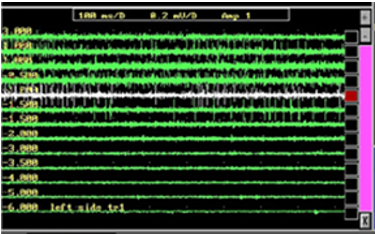
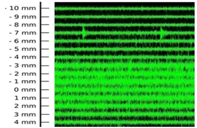
The Intra operative microelectrode recording was completed in all the 5 channels. All the 5micro electrodes were gradually passed all over the S T N and signal acquisition was done from 10 mm over to 10 mm below the S T N determined on the M R I image in a sophisticated computer parallelly networked machine. The S T N was discovered through a large-noise by a larger-baseline as well as an abnormal-discharge in conjunction with many-frequencies. The Figure 1 showing the micro electrode signal recording that was achieved from the sub thalamic nucleus.
The channel with maximum recording and the earliest recording were recorded on both sides. Intraoperative test stimulation was performed in all channels from the level at the onset of MER recording. Stimulation was done at 1mv, 3mv to assess the improvement in bradykinesia, rigidity and tremor. Appearance of dyskinesias was considered to be associated with accurate objecting. Side effects were assessed at 5mv and 7mv to ensure that the final channel chosen had maximum improvement with least side effects.
Correlation was assessed between the aspects of MER and the final channel chosen in 46 patients (92 sides).
Results
46Parkinson patients were involved in this study with their mean-age of58.1+9.1years (at the onset) with a mean disease-duration of8.8+3.64years. Because of a total of 5channels, the S T N-microelectrode neural-recordings were discovered in 3.5+1.1 on right-hemisphere-side as well as 3.6+1.04 on the left-hemisphere-side.
Conclusions
The aim of the study was to decipher the predictive-role-of intra operative neural-signals in the bilateral S T N – D B S stimulations response as well as to afford systematic and methodical-insights thru close-fitting the maximum edifying intra operative M E R signal-features and the S T N neurons and extricating the S T N – M E R signal “patterns” associated with U P D R S stage-III score -improvement. We examined the micro electrode recording through the subthalamic nucleus (S T N) high-frequency D,B,S, in Parkinson`s, documented the S T N neural-signals.
In this study, we have accomplished 80% of the variant in the scatter-plot as well as find that micro recording yields verification of correctly pointing to the micro electrode.


