Introduction
The design and development of deep brain stimulation of the sub thalamic nucleus or sub thalamic nuclei (DBS-STN) is a well established stereotactic functional neurosurgical method that eases the tremors (shaking palsy) and reinstates motor functioning in subjects i.e., patients with advanced idiopathic Parkinson's disease. However due to change of the stimulus parameters manually (i.e., in the present conventional open loop DBS devices) each year it is tailored also relatively a technological standstill. Therapeutic efficacy necessitates and also demands both surgically implantation of the DBS electrodes precisely and observant or vigilant coding or encoding the stimulus parameters. Mutually, these methods guarantee modulation of the accurate neural pathways to attain maximal remedial or restorative outcome. Nonetheless, current conventional encoding of deep brain stimuli to accomplish optimal best possible and most favorable optimal tissue establishment is a cumbersome but realistic, sensible and matter-of-fact practical procedure.1, 2
In the contemporary healthcare sectors, structural and physiologically electro-neuro approaches are existing to prospectively detect remedial or restorative establishment areas postoperatively. The two main research areas in the Parkinson disease is interventional studies (STN, GPe, i.e., MER with STN-DBS, STN-GPe) and management studies (Levodopa, i.e., L-Dopa drug trial). Computational paradigms, and prototypes for prognostics and/or diagnostics and clinical observations for diagnosis. These two paradigms or prototypes point in the direction of dorsal sub thalamic-nuclei as an effectual induced stimulus object. This area is pass through the navigate through the hyper direct pathway,3 that erstwhile vigorously implicated apprehensively in the procedures of therapeutic surgical DBS.4, 5 Establishment models or start tissue activity, specifically, have had through distinguished inputs and involvements in this field of research. While in vivo experimental dimensional gauge of tissue start or establishment is characteristically unreasonable and also unfeasible, establishment or start of tissue paradigms/ prototypes give a refined approximation of the structural brain sub-structures areas stimulated by restorative stimuli6, 7 and have reinforce hypotheses discovering the dorsal nuclei- the sub thalamic nuclei and the frenzied shortest alleyway as best and finest points of stimulus spots.8, 9 The vicinity of high β-power investigated experimented with electro-neuro-microrecording (MER) machine also have had been allied and/or connected by means of successful deep brain-stimuli and they are also recurrently concurrent through dorsal nuclei – the sub thalamic nucleus (or nuclei). 10, 11, 12 But the emergent vertebra indication and substantial verification advocates or implies that it is un-anecdote or un-intact yarn. Whilst the standard dynamic contact is located over and over again in the neighborhood of the dorsal sub thalamic nucleus perimeter, utmost at best successful induced stimulus spots are identified to fluctuate drastically or extensively transversely Parkinson`s.13, 14
Hence, it underlines that, a necessitate exist to further differentiate the 3D extent of tissue stimulated as a result of restorative induced-stimuli accurately, and traditionally that has been investigated and examined as a distinct position.15, 16, 17, 18 In the same way, numerous fluctuations of STN exterior, the four classes EEG band frequencies such as delta δ-waves (0 Hertz – 3 Hertz), theta θ-waves (4Hertz – 7Hertz), alpha α-waves (8Hertz – 12Hertz), beta β-waves (13 Hertz – 30Hertz, gamma γ-waves (31 Hertz – 200Hertz), and High-frequency waves which are above ( >200 Hertz ≤ 500Hertz, Table 1)
Table 1
Specification and classes of EEG bands frequencies δ, θ, α, β, γ and higher-band-frequency ranges
On the contrary β-band remarkably α-band, γ-band, and High-frequency fluctuations are discovered to take part in a pivotal role in Parkinson`s [25e32] plus prospectively interrelate or correlate diagnostically and prognostic ally in an evocative manners, for instance stimulus intensity and amplitude pulse-width coupling.14, 19, 20, 21 Thus, observations such as above advocates a necessitate a requirement to further construe the structural and neuro-electro physiological locus or foci of deep brain stimulus sub thalamic nuclei intrusion and involvement to comprehend better and best possible or more favorable physiological brain sub structures which are significantly important in diseased or damaged and injured STN neural points of action of Parkinson`s diseased conditions thoroughly. Hence, we analyze the STN tissue establishment and/or initiation by means of high-speed and high density electro neuro physiological microelectrode recording machine via broadband network. So we fetch these corresponding or harmonizing molds of study into a cohesive and fused study and investigation of restorative induced stimuli, by mapping microelectrode recordings to clinically-derived tissue activation models. Integrating that those personage diverse and varied an isotropy into our molds, and we get accurate subject-detailed approximation of STN movement and action.22, 23 In this study data-driven approach is also employed to categorize discover the links and relations among the vicinities of curative brain component – the STN organ element movement and action and broadband microrecording MER-features electro neuro physiologically, together with cantankerous traverse-frequency contacts or dealings and then demonstrated that MER-signal or waveform-features shall be employed to envisage or to foresee or prophecy widths of restorative establishment or start in the data set which was tested .
Broadband
Healthcare Internet of Things (I o T) mechanisms – tools and utilities devices are fetching and suitably added and famous in the engineering and technological world as countless industries- educational institutions, research and development and a myriad of innumerable organizations perceive the significance in track pathways benefit and advantage resources and observes the Parkinson subjects virtually.
Seeing that at the same time as many tools and utilities as more devices are connected with the network, industries require ensure that their network be able to capable to house, to contain over and above the supplementary elements and components at the jiffy or minute positioned to echelon suitably as additional components or elements connected.
The management and Parkinson`s depend on the allied-connected or associated components to yield the concern and heed. This reliance craving on information build the set of connections synthesized together (system) one of the mainly and nearly all imperative chunk of physical condition and strength of information technology communications in a high rich healthcare hospital environments mostly in neuroscience research centers and neurocare brain hospitals.
Materials and Methods
Discovered Parkinson`s: 107 candidates were discovered as an advanced idiopathic Parkinson`s disease candidates. All the candidates went for bilateral STN DBS brain surgery with deep brain stimulation procedure. A skilled neurosurgeon trained in stereotactic functional neurosurgery done the operation. Prior to that they were screened for cognitive and dementia issues and no one found with either cognitive dementia (CD) or cognitive impairment (CI). 16 Parkinson candidate by means of advanced idiopathic Parkinson`s disease undergone for DBS surgery for an implantation of DBS electrodes within the vicinity of sub thalamic nuclei at the NIMS hospital – a tertiary care hospital and research center for Parkinson disease and movement disorders, Hyderabad, Telangana.24 The PD candidates (Parkinson`s) were embedded bilaterally STN-DBS Medtronic micro electrodes designated with 3389 models. The micro neuro sensors (microelectrodes) were implanted with the help of STN signature patterns identified by the MER signal recording machine and also guided by the streotactic functional neuro navigation of 7 Tesla MRI. All the candidates are Parkinson`s STN DBS subjects amid constant encoding parameters and subjectively reasonable quantifiable results 180 days following the micro-neurosurgery. All the candidates had a mean-age of ±63years (SD) and a mean disease-duration of 11years. Stimulus-intensity i.e., their stimulus-amplitudes were ranged as of 1.71volts to 4.81volts with a mean of 2.84voltsAmplitude, amid stimulus-pulse-width of 60millie-seconds and 190Hertz-frequency. The three embedded electrodes were frenzied two contiguous dynamic-contacts and the rest of the electrodes were used single contact dynamically. Average DBS”OFF” and DBS”ON” with MDS-UPDRS-stage-III scores “OFF” medication were 45(19) and 31(17), with25% progress due to STN-DBS stimuli. The study was approved by the ethical board following the Helsinki principles and the written consent was obtained from all the candidates participated in this study.
Embedding Electrodes into Parkinson`s
Parkinson`s were undergone for induced deep brain stimulation subthalamic nucleus stereotactic functional frame based neurosurgery in the midst of i.e., by means of microrecording through microelectrode signal recording of STN neurons (MER signal recording of STN, i.e., MER with STN DBS). The intended designed targets were sketched and primarily consigned as of circumlocutory or circuitous target aiming, 10 millimeters tangential—lateral, 3milli meters back—posterior, and 4millie meters anterior to the mid-commissural point, by means of fine-tuning as of straight 7Tesla MRI (with luminous intensity) visualization of the ventral border of STN, on 7T-MRI (field of view ¼-200milli meters 200milli meters, 0.68x0.68,1.25 milli-meters-voxels) Medtronic, Minneapolis Minnesota, USA. MER signal recordings were done as of 10milli meters over to 5milli meters lower (±10millie meters) the projected and aimed-target scheduled on distinct-trajectory. MER signal-waveforms were acquired by the tilt of a bi polar leads, i.e., micro electrodes, augmented, and acquired – stored on to a Pentium computer machine applying custom-built Framelink program and operated in the Mat lab for the correctness and selection purposes. Neurologist detected the dorsal and ventral borders of sub thalamic nuclei in micro-neuro DBS functional neurosurgery. The then micro electrodes were embedded amid the tilt in the neighborhood of the ventral border of sub thalamic nucleus discovered according to Lozano`s electro-physiologic. Then intra operative authentication corroboration i.e., verification of the micro electrode path and electrode implantation and deep brain stimuli was done by applying a fluoro-scopy.24, 25
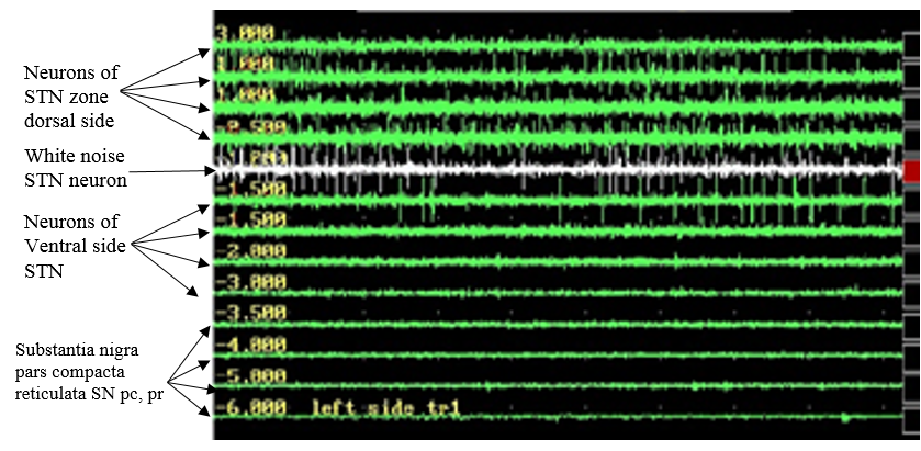
Stereo tactic aims i.e., targets were acquired by employing a specially designed sophisticated system with a stereotactic CRW frame (originally designed and modified by Cosmon-Roberts-Wells) that has a luminant MR localiser. The targeting was performed according to Lozano’s technique – 2milli meters sections are taken parallel to the plane of anterior comissure-posterior commissure line and at the level with maximum volume of red nucleus, STN is targeted at 3milli meters lateral to the antereo-lateral border of red nucleus. The co-ordinates are entered into stereo-calc software which gives the co-ordinates of the STN. Another neuro navigation software –Framelink is also used to plot the course of the electrodes and to avoid vessels. The surgery is performed with two burr holes on the two sides based on the co-ordinates. Five channels with are introduced in the midst of the central channel representing the MRI target while medial (nearer the centre) and lateral (away from the centre) are placed in the x axis while anterior(front) and posterior (back) are placed in the y axis to cover an area of 5milli meters diameter. Intra-operative recording was performed in all 5 channels. All five microelectrodes are slowly passed through the STN and recording is performed from 10milli meters above to 10milli meters below the STN calculated on the MRI. STN is identified by a high noise with a large baseline and an irregular discharge with multiple frequencies. Figure 1 shows the microelectrode recording which is obtained from sub thalamic nucleus.
Setting of deep brain stimulation electrode contacts
A highly spatio temporal resolutions by means of 10 Tesla MRI (Medtronic, Minneapolis, Minnesota, USA) scanning was accomplished 2weeks following the brain neurosurgery to envision the point of electrodes (PoE) and contacting of thee electrodes with the STN neurons with DBS stimulations. The 10 Tesla images were oversampled through lineal (linear) radial exclamation/inter-polation to equivalent the resolution of the MRI images and sloping in Talairach-space through the co muster to Talairach leaning or sloping magnetic resonance T2 weighted images by applying a mutually informative algorithmic technique in. 25 The micro electrode lead contacts were unswervingly and straightforwardly envisaged and envisioned in three dimensional imaging restorations and then the three dimensional coordinates were invoked to a program (language) meant for highly scientific and technical computing - the Mat Lab.
Modeling the Tissue Movement
Pre operative diffusion tensor imaging (D.T.I) data for each patient were acquired using a single-shot echo planar imaging sequence with a dS – S.E.N.S.E parallel-imaging scheme (reduction factor ¼ 2, field of view ¼-224milli meters 224-millimeters, 1x1-with dimension 2-millimeters-voxel`s). The diffusion weighting was encoded along sixteen16-independent orientational-directions amid a b-value of 800-sec/mille meters2. The diffusion-tensor-D.T.I images were re-sampled via cubic-spline exclamation/interpolation to equal the resolution of the resonance-images of MRI and directional in Talairach-coordinates-space via co-registration to the Talairach-directional MR images in Analysis programme. The Analysis programme-D.T.I-application was employed to compute the Eigen Values and the corresponding Eigen Values of the D.T.I images. Then the diffusion-tensors were evaluated from the Eigen values and Eigen vectors in a scientific computing tools and utilities and transformed to conductivity tensors by employing the lineal ordered radial correlation amid conductivity and diffusion-tensor Eigen values (s/d-z0.844Ssec/milliemeters3,.26 The three dimensional models of finite-element-analysis (FEA) restorative stimulations with deep-brain-stimulations’ were-built for every candidate of Parkinson`s diseased patient in another sophisticated computer programme incorporating all PD`s scan-images imaging, the induced stimulus-electrode, and clinically-determined induced stimulus parameters. Brain tissue was modeled as a block with DTI-derived an-isotropic conductivity tensors linearly interpolated onto the adaptive mesh. The electrode was modeled as an iso-tropically conductive, Medtronic-leads, model 3389,contact:1,4,107s/m;insulation:1,1013s/m27, 28 situated to equal the co-ordinates of every PD subject embedded DBS-electrode-contacts, as measured from post-operative computed axial tomography. The periphery surroundings and constraints were defined for the stimulating electrode and mass of the element i.e., mass-of-tissue. Distinctively, a stimulating-electric-potential was applied to the exterior of the dynamic reach according to automatic stimulus parameters of every PD subject, balanced or perched-floating probable’s were applied to the exteriors of the residual-contacts, and the ground-electrode was applied to the exterior of the tissue of the brain.
The finite element analysis technique was enmeshed for all PD candidates individually in the midst of better engaging, i.e., enmeshing or interconnecting applied next to the micro electrode. Induced stimulus’s then were scuttle or scamper to resolve for the stimulating voltage-potential in every-model. The models were electro static, presumptuous frequency-independent dreary-depressing-gray-matter-impedance.29 Tissue movement dimensions/sizes (TMSs) were generated for every-subject in a computer-programme by means of computing the spatial-derivative of stimulating voltage-potential30, 31 amid action and movement thresholding at the level analogous to every PD subject`s stimulus-intensity and pulse-width-amplitude amplitude.32
Microelectrode recording
The neural spikes and turning peak points of STN neurons were acquired beside the induced stimuli probe trajectory with deep brain stimulation at 0.5millemeter hiatuses, i.e., distances, straddling across as of 15 millie meters on top of 5millemeters beneath the underneath the operational target which is the sub thalamic-nuclei ventral border on the7Tesla magnetic resonance imaging by employing the neuro electro gradient.
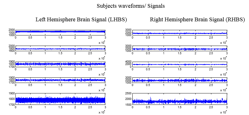
Wide band with 0.5Hertz to 15kiloHertz spiking action and movement and action field potentials, i.e., LFPs were acquired down the prod of deep brain stimuli trajectory at 0.5milliemeters A typical trajectory traverses thalamus, fields of zona-incerta, Feral/foral sub thalamic nucleus, and substantia-nigra pars compacta reticulate (SNpc, pr). Seven seconds of uninterrupted electrophysiology was recorded at each site, with the first second of recording removed from analysis to eliminate movement artifact. Each trajectory had electrophysiology recorded at 30 to 48 sites. Microelectrode recordings less than 5milliemeters from the starting-depth, which may have been affected by the cannula, were excluded from analysis. Acquired signal at each site was also visually examined for extraneous noise and excluded from analysis if at all found to be noisy. Neural-signal-data from9trajectories out of thirty-two implants were engaged or disqualified due to occurrence of noise and distortion. In total, micro electrode signals data of STNs from many spots were incorporated and integrated for the inferences deduced. Shortest prophecy of the micro electrode and the enduringly embedded macro-leads by employing intra operative-fluoro scopy established that both leads follow the same trajectory in the antereo-posterior and dorso-ventral directions. Post-operative migration of the macro stimulation lead was implicit to be negligible. By means of this association, electro physiology was spatially-mapped-to-every model-simulation down the route defined by the macro-stimulation lead of DBS envisaged on the post operative axial tomography.
At each depth, we computed spike-rate and the log-of-normalized-power in the delta:0.1e4Hertz,theta :4e8Hertz, alpha:8e13Hertz, beta:13e30Hertz, low-gamma:30e59Hertz, high-gamma:61e200Hertz,high-frequency-fluctuations:HFF;200e400, and high-frequency-bands:500e2000Hertz. Plus for main-effects, first-order interface-terms, computed as the result of every-pair of co-variates, e.g., log (beta-power) x log(low-gamma-power), were also measured throughout the diagnosis. Spike-rate was computed by high-pass-filtering, the microrecording at 300Hertz and including the number-of-threshold crosses at4.5times the waveform`s RMS. For the analysis of fluctuations, the unwanted signals of every micro-recording were projected by employing the Fast Fourier Transform (FFT). To get rid of the 50Hertz frequency-noise, the unwanted signal-values in 2.5Hertz of every 50Hertz ripples was reinstated amid the median-power in 5Hertz of the ripples.33 The system and data flow can be seen the below diagram.Figure 3
Design of classifier and validation
Predictive electrophysiological parameters for forecasting of TMA spans were identified using logistic least absolute shrinkage and selection operator (logistic LASSO). LASSO is a well-established regression method that removes uninformative covariates from linear models, thereby selecting for features that provide predictive value.34 This was carried out using MATLAB’s built-in lassoglm function. The function requires a regularization parameter, l, which determines the penalization of non-zero slopes. A parameter sweep of l was used to determine the optimal value of l to minimize divergence observed in 300-fold cross validation. The final l value used for parameter selection was one standard deviation greater than the optimal value to prevent over-fitting, following the one standard error rule for model selection.35 Parameterization of LASSO and covariate selection were performed using a training set of the data comprised of sites along 17 lead trajectories in 13-subjects (out of a total of 23implants in 16 subjects), consisting of 486 locations (spots/points).
The final classifier was a support vector machine (SVM), used due to its robustness to extreme values which are often observed in electrophysiological data. The classifier incorporated the covariates identified by logistic L. A. S. S, down the first-order terms-implicated by chosen contact-terms. The SVM was imparted training on the data-set by employing using the MATLAB built-in fitcs-vm() function, with a one standard deviation box constraint (to prevent over-fitting to outliers) and assumption of uniform prior probabili-ties. (Since there are many more sites sampled outside of thera-peutic TMAs than inside, an SVM trained on empirical prior probabilities will be biased toward classifying points as non-TMA.) Notably, the classifier analyzed each site independently, predicting whether it was inside or outside of the clinically-determined TMA, using electrophysiological features recorded at that site alone ( Figure 4A). The microelectrode recording from each site was analyzed independently of all other sites, without any spatial, trajectory, or subject information included in the analysis. Binary classifications from the SVM at each site were then spatially smoothed with a Gaussian window (s ¼ 1 millie meters) to produce a probabilistic prediction spanning the DBS lead trajectory (Figure 4B). Performance of the smoothed prediction was characterized by a receiver-operator characteristic curve.
Figure 4
A. PD Subject image Computational simulation of clinically effective tissue activation and patient MRI, B. PD Subject image Computational simulation of clinically effective tissue activation and patient MRI.

Figure 4 A. Parkinson`s model technique. Presumptions depicted in Fig B are curved above the spatio temporal regions to generate a discriminatic prophecy/guess, bookkeeping for the adjoining character of TMAs-spatially. Colors indicate probability of a site being within the therapeutic TMA, with hotter colors indicating higher probability
Figure 4 B. SVM binary predictions of clinically activated-points. Spots imagined to be within the therapeutic TMA (shown in orange) are indicated with red circles; sites predicted to be outside indicated with blue. Active contact is shown in red.
Clinico statistical investigation: the clinic-statistical/tests were accomplished by employing the scientific and technical computing Mat Lab tool. Test justification of classifier concert was verified by applying the Fisher’s t-test. The classifiers were contrasted evaluated by applying the Mc Ne mar ordeal. Performance measurements are evaluated by employing the data test.
Concert of the support-vector-machine-classifiers were resoluted by employing the hold out set-of-data encompassed of electro physiology from several sites from six-macro-stimulus-lead-trajectories in three randomly-chosen or preferred-subjects. Per se, classifier-design and corroboration were attained by exploiting a altogether part-set-of-the data which was accomplished to guarantee that classifier concert is not a upshot of over-fitting to the data of the training-instruction.
The optimized SVM classifier was compared to both a beta-only classifier and a simple STN border-based approach to targeting and coding encoding. The STN border approach assumes 2.8 V (cohort average) mono polar stimulation at the dorsal border of STN, identified in the microelectrode recording by an experienced clinical electro neuro physiologist. This passive approach to programming activates 2.0millie-meters-of-tissue in everydirection down the period of the macro leads examined in two electrode-implants with 2.8 volts mono polar induced stimuli.
Findings
The electrode implantation through deep brain stimulator is creating to contrast transversely PD-subjects, through the standard dynamic-contact situated1.89milliemeters ventral to the electro-physio-logical STN dorsal border. Tissue movement dimensions simulated/or-modeled from clinically-identified stimulus parameters of dissimilar implants period areas over, in, and/or beneath the sub thalamic nuclei. Eighteen percent of examined periods down the macro-lead were dorsal-to-nuclei, thirty-seven percent inside the dorsal-half-of-nuclei, thirty-six-percent in the ventral-half-of-nuclei, and nine-percent ventral-to-nuclei. While restorative movement points might subsist as inaccessible areas during the area of the nuclei, To defer a new prognostically practical prophecy/or-prediction of the interlude shimmering this likelihood, twofold or dual/binary-classifications of independently inferred points were smoothed by applying a Gaussian-window s¼-1milliemeters to be graded over space Figure 3 to appropriately minimize the spectra of the signals/waveforms. The smoothed predictions yielded probabilistic scores 0.90for the data-test test Figure. 5. Examination of individual trajectories in Figure 4 shows that classifier-performance can differ by insert, by a few assumptions wholly concordant by means of modeled prototype, as others exhibit insignificant overlie only.
Figure 5
The classifier concert and macro-stimulation leads of deep brain stimulation trajectories and smoothed classifier predictions, for all implants. Colors indicate probability of a site being within the therapeutic area, with hotter colors representative high likelihood. Dynamic-contact is depicted in red-color. Two-macro-leads employed two-active-contacts and the two are anodic and synchronized.

Table 2
The classy-concert.
Construal of co-variations result
Distinct-co-variations were inadequate for precisely predicting-the-effective spots-of-tissue movement down the micro surgery by applying the deep brain stimulator.
So, specific attention was the predictive-result-of-β. The β-power-plot adjacent to high frequency band Figure 3 A depicts that prominent β is a optimistic forecaster of restorative action and movement spots however in the occurrence of elevated high-frequency-β-band. Positional points by higher-β are no movement actioned by restorative stimulus whilst examined by squat higher freq band. Higher-freq. band-θ connections depict a dissimilar result: small-θ-power is a +Ve-predictor of restorative action-movement whilst concurrent by larger fh-band power 3. While high-frequency-band is a sign of sub thalamic nuclei multi unit activity, these connections point to that larger β-power and lower θ-power are optimistic assumptions of restorative action and movement in the nuclei. As the character of connections amid these fluctuations and their association is not from image examination, a huge covariance examined amid these pairs of covariates is notable Figure 7.
Figure 6
Receiver operating characteristic curve of smoothed classifier. Calculated from test data. AUC ¼ area under curve.

The prime results of connections terms-computed-plotted one versus other depict inter frequency reliance-of-predictors.
Conclusion
Analysis of the α-β contact is understandably lesser. Possibly and physio logically a singular and significant fluctuation is gathered by two α and β-bands in concordance by the larger covariance of α and β examined in the neural-data in this study, that the mold accurate for applying the contact period. Some proof /verification might be explaining insertion of the larger high-γ-frequency contact in the L. A. S. S. simulation-type-model. Nonetheless, by predicting the two-approximate places and duration of widespread and comprehensive restorative tissue therapeutic-action and movement, the modalities-presented here might be employed to rapidly approximate effectual spots and stimulus-intensities and amplitudes. Possibly and significantly further, this technique imperatively limits the stimulus-parameter-space which ought to be explored to optimize induced stimulus parameters of deep brain stimulator. In the midst of the overture of directional induced stimulus-electrodes of DBS-encoding better or superior therapeutic-windows.


