Introduction
Adaptive closed loop (ACL) approaches to surgical-therapeutic deep brain stimulation(DBS), in which neuro electro physio-logical and kinesio-logical feedback response (of the human body mechanics) is employed to acclimatize the parameters of stimuli, grip-guarantee and securefor advancing the medical management/or administration and treatment consistency of neurodegenerative diseases neuro-logical and neuro-psychic and psychiatric-disorders of Parkinson`s, minimizing induction of side-effects, and potentially plummeting the frequency of substitute surgeries for insertable pulse-generators (IPGs) by means of primary neuronal-cells. Current applications of adaptive/closed-loop approaches to surgical-DBS-therapy for reducing Parkinson’s disease (PD) have relied on sampling-potentials (the local-FPs) activity from a pair of milli-meter scale cylindrical macro electrodes along-side the lead-electrode of DBS and then utilizing computed power-spectral-features in dBs of those waveforms to establish whilst how to stimulate.1, 2 While retrofitting DBS-lead/macro electrodes for neural signal acquisition reasons erstwhile expedient for moving ACL-DBS concepts to clinical settings,3, 4 the dimensions of and spacing connecting each macro/electrode in the bipolar signal-recording pair likely under samples the spatial heterogeneity of fluctuatory sinks and sources in the object nuclei. 5, 6 Preceding reviews employing mono-polar or bi-polar electrode/or-sensor sensing technical/specifications and configurations have had noted that accustomed and customary target objects of surgical-DBS- therapy for PD, plus the subthalamic-nuclei(STN) and the global-pallidus internus (GPi)/externus (GPe), can corroborate momentous spontaneous and task-related fluctuatory activity in controls7, 8, 9, 10, 11, 12 and Parkinson`s situations.6, 8, 13, 14, 15, 16, 17, 18 Nonetheless, earlier reviews have had found that the significant inter-subject variability in the power spectral densities and specific frequency bands of such fluctuatory activity`s revealed in controls5, 18 and in primates as well.8 Such variability may possibly indeed positively stanch from phy-sio logical disparity`s in the sampled target-object amongst-subjects, however the disparity`s can echo or replicate the grade to which signal-acquisition or gathering electrode dimensions, locations, and configurations can authentically and faithfully edge the electrical-shunting of oscillationsfluctuatory-dipole motion or action movement in the areas of concern and simultaneously and selectively eliminate the prying from far-field fluctuatory-oscillations sources. This pilot-study investigated the scientific-rationale/hypothesis that DBS leads with smaller, segmented electrodes will uncover increased heterogeneity of LFP activity within basal ganglia targets of DBS therapy. Acute intraoperative human studies leveraging DBS arrays indeed suggests that oscillatory activities in the STN have a finer spatial resolution than what is detectable with commercially-available DBS leads with four cylindrical macro-electrode-contacts.13 What remains unclear is the degree to which this concept varies amongst the primary DBS targets for treating PD—that is, the STN and GPe/ GPi. Additionally, little is known concerning the degree of spatial heterogeneity of oscillatory activity in these targets across behavioral states (resting versus active) and between naïve and parkinsonian conditions. To investigate these questions, DBS arrays with electrodes segmented both along and around the lead body[26] were chronically implanted within the STN and within the GPe/GPi in two non-human primates rendered parkinsonian with systemic administration of MPTP (1-methyl-4-phenyl-1,2,3,6-tetrahydropyridine).
Primates
Primates were employed in this pilot-study. Institute ethical committee approved following Helsinki rules. The primates were endowed by ecological fortification, water, food, etc. Endeavors were put to give heed and assuage or ease any anxiety or distress for the primate’s throughout-study. The primates undergo pre - op MRI at NIMS-University by employing a reflexively protected imaging system and by applying the method showed in.
Surgical operation and implantation of DBS electrodes
Each monkey was embedded by means of a titanium head post and 2cranial opening hollows over the right hemi-sphere brain (RHB). The point-of-reference and pose-of-every cranial hollow`s were steered by a pre thera peutic-neuro-surgical clinical/prognostic neuro n-vision-navigational software-programme. The software-programme allowed three dimensional image ideas of potential leads-macro-electrodes which instill trajectories by the goal of targeting-object the GPe and GPi and/or the s-nuclei avoiding-auxiliary-trajectories in the course-of hefty-sulci, and primary-motor-cortex-ventricles. A micro drive was closed-to every hollow and applies to funnel a micro electrode (250 µicron-meter thickness, and impedance 0.8megohm-Ω –1.2megohm-Ω) in the course of an acute channel-can-nulas in to the brain. 5channel micro-electrode array-tracks were done for each target-object to find and plan the sensory motor territories of the GPe/GPi and s-nuclei. The boot-rate and patterns/or signatures of secluded STN-neurons in each of the target-objects were employed to pin point the boundaries of every nuclei-of-interest. Sensory motor territories in these nuclei were detected as those consisting neurons whose boot-rate was modul-ated-transformed by means of inert flaccid joint verbalization or volitional lobby. The site of the demonstrating, i.e., STN-neuronal signal-recording paths virtual to the cortico-spinal swathe or territory of inner pod was dogged by employing using micro stimuli, 50micro-amperes–200 micro-amperes, 300 Hertz frequency, with a duration of 0.45 seconds to inducing progress of the façade or visage, greater limit, and lesser limit and for 2primates, the global pallidus internus and externus target-object was diagrammed and embedded by a deep brain stimulation arrangement preceding to the planning and insertion of the ipsi lateral array of s-nuclei-dbs.
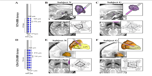
The DBS arrangement comprised thirty-two ellipsoidal that are resembling clouds microelectrodes set in 8-rows and 40-columns approximately a 600 micron-diameter shaft, amid electrode-diameters-of [three-sixty.micron×three-sixty.micron] for s-nuclei embed and [three-seventy.micron×four-seventy.micron] for the pallidus-internus/externus insert, and pivot to pivot electrode-terrain down the axis-of stimulus macro-electrode/lead was five-hundred-seventy-two micron, five-hundred-seventy-two micron for s-nuclei and pallidal internus/externus stimulus-arrays. Subsequent to the insert of the 2arrays-dbs, a computed axial tomography was also done within-the-subjects to confine the array-points in milieu of pre op imaging. The angle-direction of stimulus-array was done by employing-2-looms. Every stimulus-array assemblage had a lead-wire lengthening from the lead vertebra inside cranial-cavity-or-hollow and the assemblage provided as a fiducial for position of electrode-columns situated distal down the direct-shaft. Before insert, stimulus-arrays are scrutinized with micro scope to corroborate the arrangement. Moreover, stimuli provoked sway abbreviations or retrenchments consequentially ensuing from presumed commencement of the corti co spinal-territory of inner container were calculated. The brink current-voltages (in Amps) approximately a line of electrodes was used to identify the electrode contact(s) that most closely faced the internal cap-sule and further confirmed results from the fidu cial studym and s-nuclei stimuli array and non invasive electromyogram electrodes were positioned on the flexor aspect of forearm and bi ceps, and electromyography signals were assessed instead eye examination of establishment of muscle-fibers. Next placing of every electrode, impedance measurements (spectro-scopic) was done on every noninvasive electrode-pint. The input and output impedances in the array of ten kilo-Ohms to three hundred kilo Ohms and at 1 kilo Hertz and two-hundred kilo-Ohms to seven-hundred kilo-Ohms at twenty Hertz were estimated impractical and were excluded as of auxiliary investigation to shun the local field potentials examination.
Signal acquisition of LFPs
Te potentials were recorded simultaneously and sampled asynchronously@1.5 kilo Hertz by using α-ώ-system. LFP-signal acquisitions were positioned to a lateral titan ium cranium-skull post, which was fixed to head by ten to fifteen titan ium fillet-turns. Signals of local fields were acquired at latent condition and at voluntarily. At voluntary, dual or mutual point information-data records gathered as of deep indicators positioned on limb employing a camera(motion) and also web cam. Member sites were reconstructed by using a piece of code and the parameters extracted in a language of scientific and technical computing kine matically.
Power Spectral density
The signal to noise ratio was performed and the artifacts were removed. The frequency-spectrum for a set latent-condition and check-condition was computed through averaging with the windowing concept. Windowing was performed to avoid and to minimize spectra of the potentials. The spectral-power was computed (in decibels-dB). Filtering was performed using butter worth and chebyshev. Power spectra are computed. The spectrum shape was contrasted alongside of the point of the electrode-impedances and with correlations’< 0.05 which was statistically significant with χ2 @ 4.2857 for 1 degree of, which significant at 5% with p = 0.0425.
The field potentials in the salvage-chore were coupled to the time of movement instigation and spectro grams for every sample was computed. Average moving window was done. The z scores for the normalization is done and the z score spectral gram attained and then the SD variances σz-score of the z score spectral gram curves athwart – diagonally and the intact deep brain stimulus-array mathematically represented as
array mathematically represented as by the following equation (1)
...................................(1)
in which, N-represents the electrode number, zi(t,f) being z-score represents spectro gram, and z(t, f) mean-average z-score ‘spectro grams athwart diagonally to each and every one, z-score spectr ograms deducted to produce a disparity , z-score spectr ograms as given in (2)
Standard deviation of the spectro gram disparity is computed by the following expression 3 (mathematically represented)
.......................................(3)
The significancy of the clinico-statistic is definite at plus or minus3z-scores.
Results
The s-nuclei deep brain stimulus-array in every Parkinson was embedded down a para sagittal trajectory suchthat macro-electrode macro stimuli lead vertebra sent during the s-nuclei by electrode line ups one to five and line ups six to eight having at least one electrode within the s-nuclei – Figure 1. Histo logical corroboration of the stimulus-array places were accomplished for both, except no his to logy was obtainable. As corroboration of stimulus-array course-direction, stimuli pulsate-trains and pulse-widths 300Hertzes, 100micron-amperes to 400micron-amperes, and with a time period of 0.5seconds were carried in the course of 1 or ≥ electrodes down a sole feature of stimulus-array to discover stimulus-amplitude entry`s to extract sway jolts. Despite stimuli-array, contra lateral limb examined primarily a jolt or jerk, and then flexor aspect of fore arm and leg jerks up on inclining-up stimulus-strength. Stimuli gate for the limb typically-hand be establish to be lower-boy for elect rode links in front of inner-capsule-than for sensing-electrode links opposite absent as of inside pod for s-nuclei and pallidal array-Figure 1 and for s-nucleus-arrays, inducing electromyography-signals were evaluated in lieu-of computing a gate-voltage which is amplitude which is the strength of the signal and are with high-resolution which are giant waveforms whilst inducing by agile-line of sensing-electrode that is closure to the inner-pod.
Figure 1
Stimulations of s-nuclei and pallidal neurons with deep brain stimulator and their course-directions in Parkinsonians. Figures A and D. Stimulus-arrayscomprised of rows-and-4columnsof electrode-places amid slighter-electrode sizes and inter-electrode spacing’s of s-nuclei-dbs-arraysB and C-than pallidal-neuronal-arrays E and F. Polar plots indicate either stimulus amplitude thresholds to evoke muscle twitches E,F or stimulus-induced electromyography-voltages-peakC-while inducing through electrodes along a single-column.

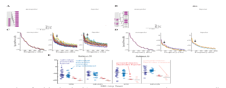
The subjects in green and Parkinsonian-conditions in which field-potential acquisitions were gathered with stimulus-arrays. These acquisitions were developed computationally and procedurally in terms of personage sensing-micro-electrodes (bipolar: adjacent row subtraction) and/or clustered hybrid macro sensing-electrodes/leads. The spectral grams by z-score complexion ruddiness (tint) show significant reductions in the strength of β-frequency-band significantly and directly prior to and later, the start of the contact-progress within the segment of bi polar acquisition-duo. Also, there was distinctly marked short γ-frequency-band:30Hertz-50Hertz action-movement instantly subsequent the start of the reach movement.
Conclusions
The findings established momentous hetero geneity in the temporal-spatio circulation of fluctuatory-oscillations action-movement in s-nuclei and in pallidal neuro nals in raw and parkinsonian`s(primates). The application of slighter fragmented stimulating microelectrodes approximately and down the stimulus-array is exposed to maximum shunting of fundamental fluctuatory-dipoles oscillations, predominantly in the β-frequency-band and in the γ-frequency-gamma-band, and ensuing within high-spectral-power and more temporal-spatio hetero geneity contrasted by bunched macro-electrode technical specifications/configurations which are unswerving by the bigger and superior cylindrical sensing-micro-electrodes employed in mainly viably accessible available deep brain stimulation devices/systems. Findings also showing that the future ACL-DBS-systems that utilize LFP feedback waveforms will strongly benefit from the use of DBS leads with smaller electrode sizes and inter electrode spacing’s. The fragmented stimulus-arras by high thickness lesser associates be capable of identifying (with better resolutions in terms of dynamic ranges) greater temporal-spatio variations in local field potentials achievement in the BG for latent condition and dynamically attaining the conditional behavior during the tasks amid raw-Parkinsonian`s. These spectral changes exhibited a spatial heterogeneity within the target nuclei that for many features could not be sensed using larger grouped macroelectrode configurations. Future development of ACL-DBS therapies that rely on sensing LFP activity will likely benefit from the use of smaller electrode sizes and interelectrode spacings that are more consistent with known anatomical subregions within target nuclei of DBS therapy.

