- Visibility 151 Views
- Downloads 27 Downloads
- DOI 10.18231/j.ijn.2020.032
-
CrossMark

Head injury related ocular manifestations: A multicenter study
Introduction
India has the rather unenviable distinction of having the highest rate of head injury in the world. In India, more than 100,000 lives are lost every year with over 1 million suffering from serious head injuries. Every day, men, women and children suffer head injuries. Head injuries can be defined as those in which there is evidence of the involvement of the brain, including concussion, loss of consciousness, posttraumatic amnesia neurological signs of brain injury, or skull fractures.[1] A trip or fall, a car accident, a sports injury – these everyday injuries can range in severity from concussion to coma. [1] About 25% of patients with head injuries will have some form of ocular manifestations and 11% of them will develop blindness. [2] Hence, the role of ocular injuries secondary to head trauma in the causation of blindness and overall prognosis of patients has become a subject of immense importance. [3]
The visual morbidity following head injuries are most often neglected and present at much later day of injury causing irreparable visual loss. Head injuries are frequently associated with ophthalmic manifestations and consequent visual morbidity, but many of the ophthalmic findings are often ignored and present much later to specialist neuro-ophthalmic clinics.[4] Hence, clinical correlation of the ophthalmic findings is important in early localization of the site of injury, better management, and improved visual prognosis of the patient with head injury. [5], [6] The aim of this study was to evaluate various ocular manifestations in cases of head injury patients, correlate them with the patients neurological status and to analyse any association between them.
Materials and Methods
The study was done as prospective multicenter based study done at three different emergency services at tertiary care centres in south India over a period of one year from June 2018-June 2019. Initial examination of all 150 patients included in the study was collection of demographic details, taking of detailed history regarding the nature of injury and the details were entered into an performa. Patients conscious level was assessed using Glasgow coma scale. The GCS includes examining patients ability to open eyes on respnse, response on verbal communication and response to motor stimulation. Revised Trauma score (RTS) included GCS, systolic blood pressure and the respiratory rate of the patient in graded the severity of head injury. Lower the score of the RTS higher the severity of the head injury and vice versa. Poorer GCS and RTS scores patients have less chances of survival. However, we tried to correlate that probability with the ophthalmic signs of neurological significance.
Patients who were uncooperative for examination were excluded from the study. Initial neurological examination of the patient was done by an on duty neurologist and later detailed examination with necessary investigations whenever needed by an neurosurgeon.
Similarly, Ophthalmological examination at initial level included assessment at bedside for any external ocular injuries with particular importance to assessment of pupillary reaction and at later level detailed slit lamp examination and dilated fundus examination. In selected patients B scan ultrasonography, X-ray orbit, CT orbit, MRI brain and orbit was done. Ocular condition or neurological condition requiring surgical intervention were treated accordingly.
Statistical Analysis
Study significance was analysed statistically by calculating p value using chi square test and fisher exact test with the help of SSPS software version 20 and a value of less than 0.05 was taken as significant.
Results
The total number of patients recruited in the study of closed head injury were 150 patients. the demographic data of the study is depicted below ([Table 1]). Out of those 150, males formed the large number with 136 patients (91%) and females were 14 (9%). The age of patients ranged from 7 to 60 and the mean age being 30.12 years. In our study it was very obvious that young adults 15-30 years were the most vulnerable population. The commonest cause for head injury in our study population being the road traffic accident. Ninety (60%) out 150 patients had history of RTA. About 33 patients (22%) had history of assault and the remaining 27 patients had history of various other kind of injuries like fall, hit by stone, injury while playing, injury by cattle ([Table 2]).
| Demographic data | N=150 |
| Sex ratio | |
| Male | 136 (91%) |
| Female | 14 (9%) |
| Age distribution | 7-60 years |
| 5-10 | 3 (2%) |
| 10-20 | 7 (4.6%) |
| 20-30 | 54 (36%) |
| 30-40 | 39 (26%) |
| 40-50 | 24 (16%) |
| 50-60 | 23(15.4%) |
| Laterality of eye involvement | |
| Right eye | 26 eyes |
| Left eye | 40 eyes |
| Both eyes | 36 eyes |
| Mode of injury | Number |
| RTA | 90 (60%) |
| Assault | 33 (22%) |
| Others (fall, hit by stone, injury while playing, injury by cattle) | 27 (18%) |
Of the 102 patients with ocular injury involvement of right eye was seen in 26 eyes, left eye in 40 eyes and both eyes in 36 eyes. The types of eye injuries in our study included, soft tissue injury of the globe and adnexae in 68 patients (45.3%), neuro-opthalmic injury in 48 patients (32%), orbital wall fracture in 30 patients (20%) and 10 patients (6.6%) had globe rupture ([Table 3]).
| Type of injury | N |
| A. Soft tissue injury | 68 (45.3%) |
| Periorbital ecchymosis | 50 (33.3%) |
| Lid laceration | 15 (10%) |
| Sub conjucntival hemorrhage | 34 (23%) |
| Proptosis | 2 (1.3%) |
| Neurogenic Ptosis | 2 (1.3%) |
| Corneoscleral tear | 10 (6.6%) |
| Hyphaema | 8 (5.3%) |
| Vitreous hemorrhage | 4 (2.6%) |
| Macular oedema | 7 (4.6%) |
| Retinal detachment | 1 (0.6%) |
| B. Orbital fracture | 30 (20%) |
| Lateral wall | 15 (10%) |
| Medial wall | 6 (4%) |
| Floor | 3 (2%) |
| Roof | 1 (0.6%) |
| C. Neuro ophthalmological deficits | 48 (32%) |
| RAPD | 30 (20%) |
| EOM restriction | 37 (24.6%) |
| Optic neuropathy | 24 (16%) |
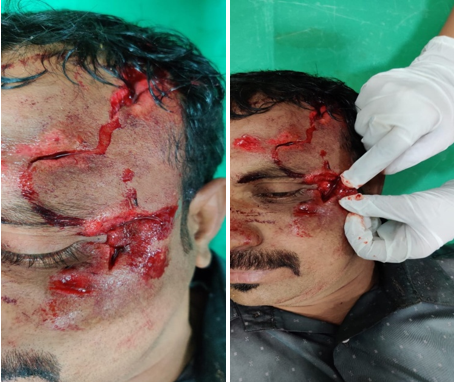
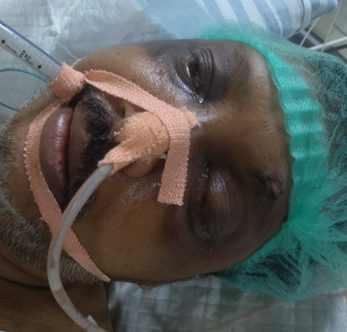
Of the soft tissue injuries the commonest type seen was peri orbital ecchymosis and oedema seen in 50 patients (33.3%), subconjunctival hemorrhage was seen in 34 patients (23%), lid laceration was seen in 15 patients (10%), corneo scleral tear was seen in 10 patients (6.6%) and macular edema was seen in 7 patients (4.6%).
The commonest of orbital wall fracture was lateral wall fracture seen in 15 patients (10%). Others included floor, superior and medial wall fractures.
The neuro-ophthalmic manifestation commonly seen was related to pupil in the form of change in shape, size and reaction (RAPD) seen in 48 patients (32%). Next extra occular movement restriction was seen in 37 patients (24.6%). Twenty four patients (16%) had some signs of traumatic optic neuropathy.
Many patients had a mixture of all of these or some of these ocular injuries. 95 patients (63.3%) of them had SCH, echymosis, orbital wall fracture, hyphaema. 22 patients (14.6%) had injuries of other organs of the body like chest, abdomen, and long bones along with head and eye. Out of 30 patients with orbital wall fracture 17 patients had associated multiple facial bone fracture ([Table 4]).
| Multiple injuries | N |
| SCH, Echymosis, orbital wall fracture, Hypheama | 95 (63.3%) |
| Chest, abdomen and long bones | 22 (14.6%) |
| Multiple facial bone fractures | 17 (11.33%) |
In our study we noticed cranial nerve palsy in 9 cases out 37 patients with EOM restriction. Sixth nerve palsy was seen in five patients, forth nerve palsy in one and third nerve palsy was seen in three patients. Rest of the patients that is 28 patients had restrictions due to local mechanical restrictions which got resolved in due course of the recovery. This association of cranial nerve palsies with restriction of EOM was significant with p value of 0.001. Out of the 48 patients with pupil involvement 30 patients had various grades of RAPD and remaining 18 patients had altered pupil shape due to sphincter tear. Vision in patients with RAPD ranged from light perception to 20/20. Six patients with severe RAPD showed fundus changes of complete optic atrophy in due course. When relation between RAPD and vision of patients was assessed 6 patients out 30 patients with RAPD had vision less than 20/200 in comaprision to 20 patients without RAPD, this numbers had a p value of 0.78 which was not statistically significant. 6 patients with RAPD >grade 2 had vision <20/200 in comparison to 0 patients with RAPD <grade 2. This number was statistically significant with p value of 0.0004 ([Table 5]).
Patients who had GCS of less than 10 were also assessed to the changes in pupil reaction. 18 patients had GCS of less than 10 and all 18 patients had associated pupillary manifestation. This number indicated significant association between head injury and pupil involvement.
| Correlation of Cranial nerve palsy and its effect on extra ocular movements | ||||
| Intact CN | Damaged CN | Total | P value | |
| Full EOM | 65 | 0 | 65 | 0.001 |
| Restricted EOM | 28 | 9 | 37 | |
| Total | 93 | 9 | 102 | |
| Correlation between RAPD and vision | ||||
| Vision <20/200 | Vision >20/200 | Total | P value | |
| RAPD+ | 6 | 24 | 30 | 0.78 |
| RAPD- | 20 | 100 | 120 | |
| 26 | 124 | 150 | ||
| Correlation between severity of RAPD and vision | ||||
| Vision <20/200 | Vision >20/200 | Total | P value | |
| < grade 2 | 0 | 20 | 20 | 0.0004 |
| >grade 2 | 6 | 4 | 10 | |
| 6 | 24 | 30 | ||
| Correlation between pupil involvement and severe head injury with GCS<10 | ||||
| GCS <10 N=18 | GCS 10-15 N=132 | |||
| Pupil involvement | 18 | 30 |
| Signs | N=70 | No. of deaths N=24 |
| RAPD | 30 | 10 |
| Papilloedema | 9 | 5 |
| Sixth nerve palsy | 5 | 2 |
| Neurogenic ptosis | 2 | None |
| Traumatic optic neuropathy | 24 | 7 |
The next part of our study was to correlate ocular findings to severity of head injury by comparing them to GCS and RTS scores [[Table 7]]. Mild head injury patients (GCS of 13–15 and RTS of 10–12) were 110 (73%) and out of them 64 patients had eye involvement and 10 patients had neurological involvement. None of these patients died. Moderate head injury (GCS 11–12), were 22 patients (15%) of which 20 patients had eye related manifestations. Among these all 20 patients had significant neurological deficit and 6 patients died. Severe head injury patients were 18 in number of which all 18 patients had neurological eye signs and all of them died.
| GCS | RTS | Cases of head injury N=150 | Cases of ocular injury N=102 | Patients with neuro ophthamic signs N=48 | Final status of patient |
| 3-5 | 1-3 | 2 | 2 | 2 | 2 deaths |
| 6-8 | 4-5 | 6 | 6 | 6 | 6 deaths |
| 9-10 | 6-7 | 10 | 10 | 10 | 10 deaths |
| 11-12 | 8-9 | 22 | 20 | 20 | 6 deaths |
| 13-15 | 10-12 | 110 | 64 | 10 | No deaths |
There was a significant correlation of the GCS, neurodeficit, and the ocular signs with outcome [[Table 8]]. Pupillary abnormalities, optic nerve injury, and lateral rectus palsy pointed toward a poorer outcome. All the patients who died had ocular involvement of neurological significance. The outcome is worse in patients with GCS 6–8 with ocular involvement and neurodeficit (P 0.0026 and P 0.01 respectively, Fisher’s exact test) and those with GCS head injury (P = 0.0003, Fisher’s exact test). The GCS, neurodeficit, and ocular signs contribute significantly to the prediction of outcome.
| Criteria | Outcome of death p value |
| GCS head injury vs death | |
| 3-8 | 0.0003 |
| 9-15 | |
| GCS ocular involvement vs deaths | |
| 3-8 | 0.0026 |
| 9-15 | |
| GCS neuro deficit vs deaths | |
| 3-8 | 0.01 |
| 9-15 |
Discussion
India being one of the developing country which is fast adopting to cosmopolitan culture, RTA will play a large role in physical suffering of young population. Recent stats say that India accounts for roughly about 10% of global RTA cases.[4] RTAs were the most common cause of head injury due to high‑velocity impact. Other studies also showed almost similar observations. [7], [8] Raju reported 47.5% of cases because of RTA and 32.5% of cases due to fall from height. [9] Similar statistical data was found in our study were RTA accounted for 90% of the head injury patients.
This study was done on 150 head injury patients admitted in neuro surgery wards with ophthalmic findings. Age of the patients ranged from 7 to 60 years with a mean of 30.12 years (±8.78 years). Young adult males (15–30 years) were the major group who sustained head injury, i.e., 100 of the 150 of head injury cases. Our study finding is similar to the findings in other studies, for example, Kulkarni et al. [7] showed that the young adult males (21–30 years) were more vulnerable. Odebode et al. [8] showed a peak during third decades (21– 30 years) of life. Sharma et al. [10] showed a peak during 21–40 years. This vulnerability of the young is due to the increased association with outdoor activities.
None of the patients had diplopia, or nystagmus in our study. Diplopia is a finding frequently encountered in head injury patients. In our study 33% of patients had periorbital echymosis, 23% patients had sub conjunctival hemorrhage and 32% patients had pupilary manifestations. These findings were comparable to other studies. [7], [8], [10] Among the orbital wall fracture lateral wall fracture is the commonest fracture in our study which may be attributed to the mechanism of impact during RTA where lateral wall gets injured most of the time on verge of protecting the eyeball. [11]
In our study there were nine patients with associated cranial nerve palsy who also had EOM restriction and the association is statistically significant indicating causation of EOM restriction due cranial nerve injury in head injury patients. Moster et al [12] reported cranial nerve injuries with associated ocular signs in head injury patients. Mariak et al 14 reported serious cranial nerve damage in 12 patients subjected to brain autopsy after death.
Examination of pupil for its size, shape and its reaction to light plays very important role in initial examination of patient with head injury. In our study 100% of patients with GCS <10 that is all 18 patients had abnormal pupil indicating very significant association between pupillary involvement and head injury. Thirty patients had signs of RAPD and out of them six patients had vision less than 20/200. This number did not reach statistical significance (p value 0.78) though patients with grade 2 and above RAPD had significant vision deterioration (p value 0.004). International trauma study 15 reported similar pupillary findings in head injury patients and reported vision recovery in patients with pupillary abnormalities following steroid injection as treatment.
Our study reports a statistically significant association between GCS in all head injury patients to number of deaths (p value 0.0003), GCS in ocular involvement patients and number of deaths (p value 0.0026), GCS in neuro ophthalmic deficits and number of deaths (p value 0.01). In a study by Masila et al. [13] a positive correlation was seen between severe head injury (GCS < 8) and occurrence of ocular signs.
Our study had few drawbacks too which might have affected the final outcome like assessment of ocular motility was not possible in patients under coma as few signs like third nerve misdirection arise after sometime following head injury. Test like Visually Evoked Potential concerned to optic nerve function in picking up subtle optic nerve injury was not performed in few patients due to lake of availability in certain circumstances or due to financial constraints from patients side.
Conclusion
Our study stresses again on the importance of complete ophthalmological examination at the earliest in association with Glasgow coma scale in patients with head injury to assess the final outcome of the injury. Ophthalmological examination also acts as a prognostic marker hence should be repeated at regular intervals to monitor the signs of deterioration as well as recovery. Our study hence emphasizes on importance of incorporating ophthalmic assessment into routine head injury assessment.
Source of Funding
None.
Conflict of Interest
None.
References
- J. F. Annegers, J. D. Grabow, L. T. Kurland, E. R. Laws. The incidence, causes, and secular trends of head trauma in Olmsted County, Minnesota, 1935-1974. Neurol 1980. [Google Scholar]
- GVS Murthy, M Chandra, SK Gupta, P Vashist, M Gogoi, S Vats. Epidemiological study of ocular trauma in an urban slum population in Delhi, India. Indian J Ophthalmol 2008. [Google Scholar]
- Robert S. Baker, Avrom D. Epstein. Ocular motor abnormalities from head trauma. Surv Ophthalmol 1991. [Google Scholar]
- D J Thurman, L Jeppson, C L Burnett. Surveillance of traumatic brain injury in Utah. West J Med 1996. [Google Scholar]
- J Hutchison. Four lectures on compression of the brain. Clin Lect Rep Med SurgStaff Lond Hosp 18671868. [Google Scholar]
- L Kowal. Ophthalmic manifestations of head injury. Aust N Z J Ophthalmol 1992. [Google Scholar]
- A R Kulkarni, S P Aggarwal, R R Kulkarni, M D Deshpande, P B Walimbe, A S Labhsetwar. Ocular manifestations of head injury: a clinical study. Eye 2005. [Google Scholar]
- T O Odebode, D S Ademola-Popoola, T A Ojo, A A Ayanniyi. Ocular and visual complications of head injury. Eye 2005. [Google Scholar]
- N Raju. Ocular manifestations in head injuries. Indian J Ophthalmol 1983. [Google Scholar]
- Bhavana Sharma, Rachna Gupta, Reena Anand, Rashmi Ingle. Ocular manifestations of head injury and incidence of post-traumatic ocular motor nerve involvement in cases of head injury: a clinical review. Int Ophthalmol 2014. [Google Scholar]
- Roberto Becelli, Giancarlo Renzi, Maurizio Perugini, Giorgio Iannetti. Craniofacial Traumas: Immediate and Delayed Treatment. J Craniofac Surg 2000. [Google Scholar]
- M Moster, N J Volpe, M S Kresloff. Neuro-ophthalmic findings in head injury. Neurol 1999. [Google Scholar]
- F Masila, J Kiboi, S Marco, M Njuguna. Ocular findings in patients with head injury. J Ophthalmol East Cent S Afr 2014. [Google Scholar]