- Visibility 55 Views
- Downloads 10 Downloads
- DOI 10.18231/j.ijn.2024.046
-
CrossMark

Ventriculoperitoneal shunt associated meningitis caused by Globicatella sanguinis: Review of an emerging human pathogen
Introduction
The genus Globicatella has two species sanguinis and sulfidifaciens. Globicatella sanguinis is an unusual and rare pathogen causing bacteraemia, meningitis, and urinary tract infection in humans.[1], [2] Several unidentified streptococcus-like clinical isolates were characterised in the USA in 1992 and G. sanguinis was first described as a new genus and species of catalase-negative, facultatively anaerobic, non-motile, alpha-hemolytic, Gram-positive cocci (GPC) forming short chains or pairs by Whitman, et al.[3] This organism has also been isolated from animals with a report of G. sanguinis isolated from a lamb with meningoencephalitis in Spain.[4] The other species, G.sulfidifaciens was first described in 2001, with documented isolation only from animals. It has a 99.2% similarity in 16S rRNA gene sequencing to G. sanguinis, but is different in biochemical reaction.[5] Due to its colonial morphology, this pathogen could be readily misidentified with Streptococcus pneumoniae or viridans streptococci 7 leading to an underestimation of Globicatella infections. The epidemiology and clinical significance of this pathogen remain largely unkown as Globicatella spp. are rarely isolated from clinical samples.
Case Report
We present a case of a 36 yr old male patient who suffered a RTA back in 2013. Left Frontotemporoparietal decompression and craniectomy was done. The post traumatic hydrocephalus was managed by placement of a V.P shunt followed by cranioplasty. He was discharged with a healthy shunt and advised for routine treatment and physiotherapy. In May 2022, he was again admitted with an episode of seizure and chest wall abscess for which Incision and drainage was undertaken. Eventually he was discharged with antibiotic coverage (Tab Augmentin and clindamycin). In June 2022 he presented again to the emergency with pus discharge from the shunt tract, vomiting and fever. His vitals were stable, Glassgow coma scale-Eye opening-4, Verbal -5, Motor-6, Pulse Rate-72bpm, Respiratory Rate-16/min, Blood Pressure-130/80mm/Hg. Local examination revealed left chest wall sinus indurated with active CSF leak with pus flakes. There was no history of loss of consciousness or ENT bleed. As the shunt was infected a shunt exteriorisation was planned with external ventricular drainage and a provisional diagnosis of shunt associated meningitis was made. The patient was started on empirical antibiotics. (Injection amikacin and cefoperazone -sulbactam) but the patient continued having spikes of fever.
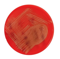
A lumbar puncture was performed and CSF sent for culture. The CSF sample was subcultured on blood agar, chocolate agar and MacConkey agar. There was no growth in the MacConkey agar. The blood and chocolate agar both showed a faint growth of tiny rough alpha haemolytic colonies after overnight incubation (24hrs) at 370C with 5% CO2.([Figure 1]) These colonies became more pronounced after 48 hrs of incubation. The colonies were catalase negative and yielded gram-positive cocci in chains on gram stain. Theses colonies were identified as Globicatella sanguinis by Vitek 2 systems and was further confirmed by MALDI-TOF MS. Antibiotic Susceptibility Testing was performed by both Vitek 2 and Kirby bauer disc diffusion method according to CLSI standards for streptococcus spp. The isolate was found to be sensitive only to Vancomycin and Linezolid and resistant to Penicillin, Ampicillin, Cefotaxime, Ceftriaxone, Levofloxacin and Clindamycin. The patient was started on Linezolid and showed a subsidence of fever and resolving of symptoms. Prior to shunt replacement, a CSF was sent again for culture which came out to be sterile. After negative CSF culture, a definitive ventriculoperitoneal shunt was implanted.
Discussion
A search of all the case reports of human infections due to this pathogen in all published medical literature available on PubMed and Google Scholar (all years until July 2022) was done, and all references were retrieved and reviewed.
The entire published clinical experience with Globicatella infections (50 cases) is summarized in the[Table 1].
As seen from the above table this pathogen as causative agent of infection has been documented since about 50 years. There are about 50 cases reported with this infection. This infection showed a female preponderance with 33 (73%) females and 12 (27%) males. Maximum number of cases were found in the extremes of age-group. There were 9 (27.5%) cases in the age group of 80-90 years followed by 8 (20%) cases in the 0 -10 years age group. Maximum cases were reported from U.S.A (n=25) followed by Canada. Three cases were reported each from Denmark, Germany and India. Out of the reported 50 cases, maximum isolation was from blood samples (n=33, 58%) followed by CSF (n=9, 18%). Most of the cases were associated with clearing of the infection, only 2 cases died. One was a 5-month-old baby with an underlying condition of posterior fossa tumour and the other was a 80 year female who had chronic diarrhoea and was a known case of diabetes mellites. Identification was done in maximum cases by Rapid ID 32 Strep (n=27, 64%). 8 cases were diagnosed by 16 S r RNA sequencing, 5 cases were diagnosed by MALDI-TOF.
Though its role as a human pathogen is confirmed, it still remains only partially known because of the trouble involved in identification and the small number of reports pertaining to Globicatella. Furthermore, it has been demonstrated that Globicatella spp. are commensal organisms in humans which makes it even more difficult to eatablish it as a cause of disease. [6]
|
Year of publication |
Place |
Reference number |
Author |
Gender |
Patient age |
Underlying condition |
Site of isolation |
Identification |
Treatment |
Outcome |
|
1994 – 2000 |
US - 22 Canada - 5 |
Shewmaker |
M - 6 F - 17 NA - 4 |
0-3 yrs - 5 25-50 yrs - 2 50-75 yrs - 3 75+ yrs – 8 NA - 9 |
NA |
Blood - 21 CSF – 2 Urine - 4 |
Rapid ID 32 strep - 27 |
NA |
NA |
|
|
2006 |
Taiwan |
Lau sk |
F |
80 |
Chronic diarrhoea, Diabetes Mellitus |
Blood |
16S rRNA sequencing |
NA |
Death |
|
|
2006 |
Taiwan |
Lau sk |
F |
92 |
Dementia, Chronic heart faliure |
Blood |
16S rRNA sequencing |
CXM.CAZ |
Alive |
|
|
2007 |
Denmark |
Abdul-Redha |
F |
23 |
Intravenous drug use, Right-sided endocarditis, Hepatitis C |
Blood |
Rapid ID 32 strep, partial 16S rRNA sequencing |
CXM, PN |
Alive |
|
|
2007 |
Denmark |
Abdul-Redha |
F |
56 |
Alzheimer's disease, Hypertension |
Blood |
Rapid ID 32 strep, partial 16S rRNA sequencing |
PN |
Alive |
|
|
2007 |
Denmark |
Abdul-Redha |
M |
82 |
Crohn's disease, Atrial fibrillation |
Blood |
Rapid ID 32 strep, partial 16S rRNA sequencing |
CXM |
Alive |
|
|
2007 |
Germany |
Seegmuller |
F |
69 |
VP Shunt |
CSF |
Rapid ID 32 strep, Phoenix pmic/id-56 |
CTRX |
Alive |
|
|
2010 |
France |
Hery-Arnaud |
F |
56 |
NA |
CSF |
16S rRNA sequencing |
CTX, FOS |
Alive |
|
|
2012 |
India |
Jain N |
M |
70 |
Craniectomy |
CSF |
Vitek 2 |
VA, LEVO |
Alive |
|
|
2012 |
Japan |
Matsunami |
M |
94 |
Dementia, CHF, Nephrolithiasis |
Blood |
16S rRNA sequencing |
A/S, VA |
Alive |
|
|
2012 |
NA |
Aseefa S |
M |
63 |
Mixed myelodysplastic and myeloproliferative disease, Chronic renal insufficiency, Obstructive uropathy with B/L ureteral stent |
CSF |
NA |
NA |
NA |
|
|
2016 |
Korea |
hs yang |
M |
85 |
Parkinson's disease, Asthma, Hypertension, staying at nursing home |
Blood |
Partial 16S rRNA sequencing |
VA, CTRX |
Alive |
|
|
2016 |
Assam, India |
Devi U |
F |
2 D |
None |
CSF |
16S rRNA sequencies |
CLOXACILLIN AK |
Alive |
|
|
2016 |
Japan |
s kurogi |
F |
80 |
Colon cancer, Brain stroke, Dementia, Hypertension |
Urine |
16S rRNA sequencing |
A |
Alive |
|
|
2017 |
New York |
Miller AO |
F |
72 |
Obesity, Gastric lap banding, Tobacco |
Hip synovium |
Partial 16S rRNA Sequencing, MALDI-TOF-MS |
VA |
Alive |
|
|
2017 |
New York |
Miller AO |
F |
54 |
Obesity, Diabetes Mellitus, Gastric bypass, Tobacco |
Blood |
MALDI-TOF-MS |
LZ |
Alive |
|
|
2017 |
Turkey |
Atkas E |
F |
43 |
Diabetic nephropathy on hemodialysis- femoral cathteter infection |
Blood |
16S rDNA sequencing |
VA |
Alive |
|
|
2018 |
Japan |
Takahasi |
F |
87 |
Endocarditis following UTI |
Blood |
Rapid ID 32 Strep,16S rRNA sequencing |
AMPICILLIN |
Alive |
|
|
2018 |
Korea |
Ahn K |
F |
76 |
Hypertension and Degenerative arthritis |
Blood |
16 S rRNA sequencing |
VA AND LEVO |
Alive |
|
|
2018 |
New York |
S,sangli |
F |
64 |
Hypertension and Proctocolitis |
Blood |
Vitek |
BROAD APECTRUM ANTIBIOTICS |
Alive |
|
|
2019 |
Turkey |
Hasbek |
F |
39 |
Lumboperitoneal shunt |
CSF |
Maldi biotyper 2.3, 16 S rDNA sequence analysis |
NA |
NA |
|
|
2022 |
Morocco |
Skali H |
F |
5 M |
Posterior fossa tumor |
CSF |
MALDI-TOF -MS |
NA |
Death |
|
|
2022 |
India |
Gupta B |
M |
9 |
Corneal abscess and Endophthalmitis |
Pus |
MALDITOF-MS and VITEK 2 |
NA |
NA |
|
|
2022 |
Burkina faso |
Kabore |
NA |
NA |
3 Acute cellulitis 1 Chronic cellulitis 1Apical periodonitis |
Pus |
API 20 strep gallery (biomérieux, france) |
NA |
NA |
Despite the similarity Globicatella has with other Gram positives, a few distinct differentiating characters exist, such as cellular arrangement of the cells in the Gram stain, Globicatella forms chains while the aerococci form tetrads and clusters. Biochemical tests that can help in identification of this pathogen include negative leucine aminopeptidase reaction (LAP) and growth in the presence of 6.5% NaCl. The viridans streptococci are pyrridonylarylamidase (PYR) negative and LAP positive and do not grow in the presence of 6.5% NaCl. Susceptibility to the third-generation cephalosporins is also different: G. sanguinis is resistant while group A streptococcus is susceptible. [7]
After the extensive literature search till date, we believe that this is the first case of VP shunt associated meningitis in a post trauma patient due to this pathogen. See gmuller and coulleges also presented a similar case of VP shunt associated meningitis due to this rare pathogen but the patient had no history of trauma and the shunt was placed to manage a decompensated hydrocephalus.[9] The second case had meningitis due to infection in the lumbo peritoneal shunt on day 10 th of hospitalisation. [20]
16S rRNA gene sequencing continues to be the best tool in the characterisation of rarely encountered bacteria and defining their clinical significance. But, in most of the clinical laboratory, MALDI-TOF has emerged as a useful tool. The mass spectrometry has the potential of being an accurate tool for catalase negative gram-positive cocci identification even for species with difficult diagnosis. [24]
With the increasing number of device implementations, medical procedures and increased survival length of patients living with advanced grades of immunosuppression, more case reports with rare pathogens have come to light. Therefore, increasing the diagnostic power of clinical microbiology laboratories by molecular methods and renewal of the databases of commercial identification systems are proving to be of utmost importance for control of infections caused by such rare pathogens.
Conclusion
Due to scarcity of literature, the epidemiology and the clinical significance of this pathogen remains largely unknown. Due to greater numbers of isolation of this rare species, the clinicians understanding about its clinical significance and antibiotic susceptibility also needs to be expanded.
Source of Funding
None.
Conflict of Interest
None.
References
- SK Lau, PC Woo, NK Li, JL Teng, KW Leung, KH Ng. Globicatella bacteraemia identified by 16S ribosomal RNA gene sequencing. J Clin Pathol 2006. [Google Scholar]
- N Jain, P Mathur, MC Misra. Globicatella sanguinis meningitis in a post head trauma patient: first case report from Asia. J Infect Dev Ctries 2012. [Google Scholar]
- WB Whitman, BM Trust. . Bergey's manual of systematics of archaea and bacteria 2015. [Google Scholar]
- AI Vela, E Fernández, A Las Heras, PA Lawson, L Domínguez, MD Collins. Meningoencephalitis associated with Globicatella sanguinis infection in lambs. J Clin Microbiol 2000. [Google Scholar]
- P Vandamme, J Hommez, C Snauwaert, B Hoste, I Cleenwerck, K Lefebvre. Globicatella sulfidifaciens sp. nov., isolated from purulent infections in domestic animals. Int J Syst Evol Microbiol 2001. [Google Scholar]
- G Héry-Arnaud, A Doloy, S Ansart, GL Lay, AL Flèche-Matéos, R Seizeur. Globicatella sanguinis meningitis associated with human carriage. J Clin Microbiol 2010. [Google Scholar]
- S Sangli, A Thakkar, S Noronha, V Santibanez, S Chakupurakal, G Narayanswami. Septic Shock Due to Globicatella Sanguinis Bacteremia. Am J Resp Crit Care Med 2018. [Google Scholar]
- RJ Abdul-Redha, U Balslew, JJ Christensen, M Kemp. Globicatella sanguinis bacteraemia identified by partial 16S rRNA gene sequencing. Scand. Scand J Infect Dis 2007. [Google Scholar]
- M Matusnami, M Sogi, H Kitazono, N Hosokawa, Y Otsuka, K Ohkusu. Urosepsis caused by Globicatella sanguinis and Corynebacterium riegelii in an adult: case report and literature review. J Infect Chemother 2012. [Google Scholar]
- S Assefa, F Rezai, M Delman, K Fless, M Litinski, P Yodice. Globicatella sanguinis meningoencephalitis and acute brain abscesses in an immunocompromised patient. Crit Care Med 20121. [Google Scholar]
- HS Yang, YJ Kim, MS Lee, HJ Lee. Globicatella sanguinis Bacteremia in a Non-Immunocompromised Patient Identified by 16S rRNA Gene Sequencing: First Case in Korea. Clin. Lab 2016. [Google Scholar]
- U Devi, R Bora, V Malik, J Mahanta. Isolation of globicatella sanguinis from cerebrospinal fluid of a neonate. Int J Biol Med Res 2016. [Google Scholar]
- AO Miller, SP Buckwalter, MW Henry, F Wu, KF Maloney, BK Abraham. Globicatella sanguinis Osteomyelitis and Bacteremia: Review of an Emerging Human Pathogen with an Expanding Spectrum of Disease. Open Forum Infect Dis 2017. [Google Scholar] [Crossref]
- E Aktaş, NC Gürsoy, T Sakacı, Y Koc, AA Hamidi, E Bulut. Femoral hemodialysis catheter-related bacteremia due to Globicatella sanguinis: challenges in species identification. Mikrobiyol Bul 2017. [Google Scholar]
- S Takahashi, C Xu, T Sakai, K Fujii, M Nakamura. Infective endocarditis following urinary tract infection caused by Globicatella sanguinis. IDCases 2018. [Google Scholar] [Crossref]
- K Ahn, GY Hwang, KJ Yoon, Y Uh. Globicatella sanguinis bacteremia in a Korean patient. Ann Clin Microbiol 2018. [Google Scholar]
- I Seegmuller, M Van Der Linden, C Heeg, RR Reinert. Globicatella sanguinis is an etiological agent of ventriculoperitoneal shunt-associated meningitis. J Clin Microbiol 2007. [Google Scholar]
- M Hasbek, Ü Özüm. A Case of Meningitis Caused by Globicatella sanguinis in a Patient with a Lumbo-peritoneal Shunt. Mikrobiyol Bul 2019. [Google Scholar]
- H Skali, AL Hanchi, M Laghmari, H Ghannane, SA Benali, N Soraa. A Case Report of Pediatric Bacterial Meningitis due to the Rare Isolate, Globicatella Sanguinis. Sch J Med Case Rep 2022. [Google Scholar]
- B Gupta, AK Jain, M Saini, M Sardana, R Soni, A Angrup. Globicatella sanguinis corneal abscess with endophthalmitis. JAAPOS 2022. [Google Scholar]
- WA Kaboré, SR Dembélé, N Barro. Detection, characterization, and antimicrobial susceptibility of Globicatella sanguinis isolated from endodontic infections in Ouagadougou, Burkina Faso. Burkina Faso Turk Endod J (TEJ) 2022. [Google Scholar]
- PL Shewmaker, AG Steigerwalt, L Shealey, R Weyant, RR Facklam. DNA relatedness, phenotypic characteristics, and antimicrobial susceptibilities of Globicatella sanguinis strains. J Clin Microbiol 2001. [Google Scholar]
- S Kurogi. A case of urinary tract infection caused by Globicatella sanguinis. J Jpn Soc Clin Microbiol 2016. [Google Scholar]
- M Almuzara, C Barberis, VR Velázquez, MS Ramirez, A Famiglietti, C Vay. Matrix-assisted Laser desorption ionization-timeof-flight mass spectrometry (MALDI-TOF MS) as a reliable tool to identify species of catalase negative gram-positive cocci not belonging to the streptococcus genus. Open Microbiol J 2016. [Google Scholar] [Crossref]